. 2020 Dec 4:16:2983-2998.
doi: 10.3762/bjoc.16.248. eCollection 2020.

Secondary metabolites of Bacillus subtilis impact the assembly of soil-derived semisynthetic bacterial communities

Affiliations

Secondary metabolites of Bacillus subtilis impact the assembly of soil-derived semisynthetic bacterial communities

Heiko T Kiesewalter et al. Beilstein J Org Chem. .

Abstract

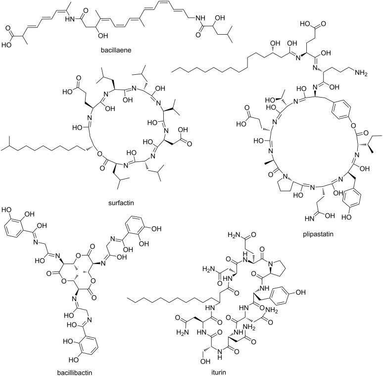
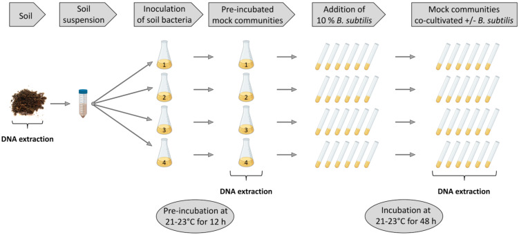
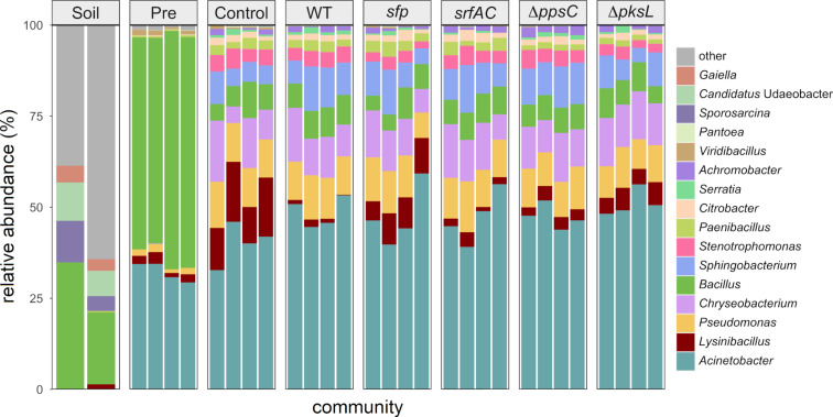
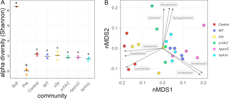
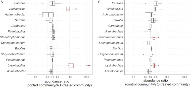
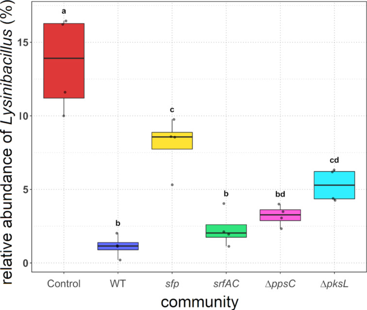
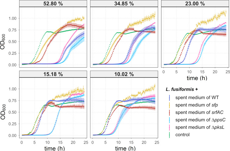
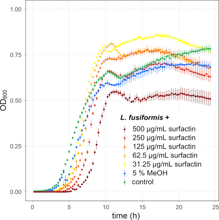
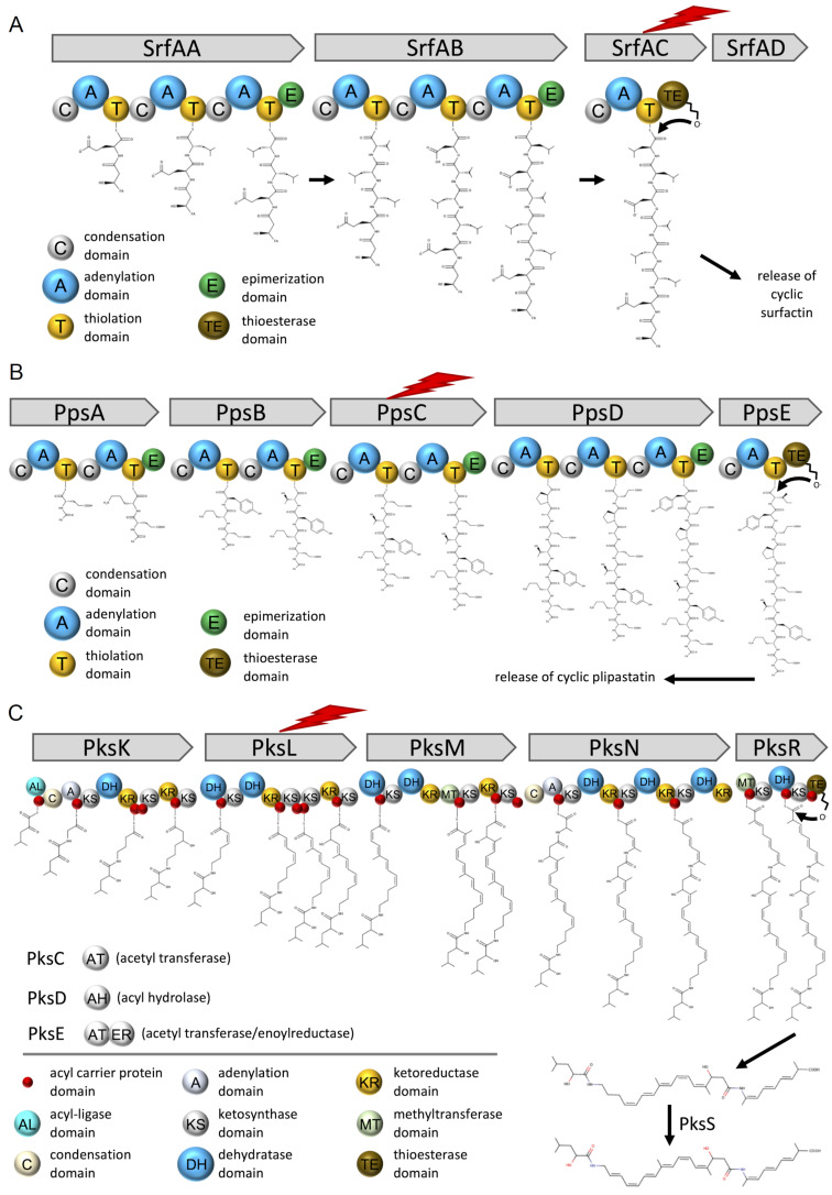
Secondary metabolites provide Bacillus subtilis with increased competitiveness towards other microorganisms. In particular, nonribosomal peptides (NRPs) have an enormous antimicrobial potential by causing cell lysis, perforation of fungal membranes, enzyme inhibition, or disruption of bacterial protein synthesis. This knowledge was primarily acquired in vitro when B. subtilis was competing with other microbial monocultures. However, our understanding of the true ecological role of these small molecules is limited. In this study, we have established soil-derived semisynthetic mock communities containing 13 main genera and supplemented them with B. subtilis P5_B1 WT, the NRP-deficient strain sfp, or single-NRP mutants incapable of producing surfactin, plipastatin, or bacillaene. Through 16S amplicon sequencing, it was revealed that the invasion of NRP-producing B. subtilis strains had no major impact on the bacterial communities. Still, the abundance of the two genera Lysinibacillus and Viridibacillus was reduced. Interestingly, this effect was diminished in communities supplemented with the NRP-deficient strain. Growth profiling of Lysinibacillus fusiformis M5 exposed to either spent media of the B. subtilis strains or pure surfactin indicated the sensitivity of this strain towards the biosurfactant surfactin. Our study provides a more in-depth insight into the influence of B. subtilis NRPs on semisynthetic bacterial communities and helps to understand their ecological role.

Keywords: Bacillus subtilis; Lysinibacillus fusiformis; bacterial community; chemical ecology; nonribosomal peptides; surfactin.


Figures

Figure 1
Figure 1
Overview of the NRPs surfactin, plipastatin, bacillibactin, and iturin as well as the hybrid NRP-PK bacillaene produced by Bacilli.
Figure 2
Figure 2
Overview of the experimental setup. A soil suspension, obtained from a soil sample, was used as an inoculum for four independent replicates and preincubated for 12 h. Enriched precultures were aliquoted and supplemented with 10% B. subtilis strains or left untreated and incubated for 48 h. DNA was extracted from the soil sample, preincubated soil suspensions, and mock communities. Parts of this figure were created using BioRender.com.
Figure 3
Figure 3
The taxonomic summaries are showing the relative abundance of the most abundant genera for each replicate of the soil sample (“soil”), 12 h preincubated soil suspensions (“Pre”), and untreated (“Control”) or treated mock communities with either B. subtilis wild type (“WT”), the NRP-deficient strain sfp, the surfactin mutant srfAC, the plipastatin mutant ∆ppsC, or the bacillaene mutant ∆pksL, cocultivated for 48 h. Genera are classified as “other” when the relative abundance is <2% (“Soil”), <1% (“Pre”), or <0.19% (in all differently treated mock communities).
Figure 4
Figure 4
Diversity analyses of the soil sample (“Soil”), 12 h preincubated soil suspensions (“Pre”), and untreated (“Control”) or treated mock communities with either B. subtilis wild type (“WT”), the NRP-deficient strain sfp, the surfactin mutant srfAC, the plipastatin mutant ∆ppsC, or the bacillaene mutant ∆pksL, cocultivated for 48 h. A) Alpha diversity (in Shannon) of the different samples. Each point represents a replicate, while the line indicates the mean of the Shannon diversity indexes. B) Beta diversity of the mock communities calculated with the Bray–Curtis dissimilarity and visualised as circles in a nMDS. The vectors, each labelled with the corresponding genus, represent the ASVs, with the highest correlating with the nMDS ordination. The vector lengths are proportional to the level of correlation.
Figure 5
Figure 5
Abundance ratios for each genus and replicate (points) in the control community compared to the WT-treated (A) and to the sfp-treated community (B). Red-box plots highlight the statistical significance, which is defined as P ≤ 0.05 (*), P ≤ 0.01 (**), and P ≤ 0.001 (***).
Figure 6
Figure 6
The relative abundance of Lysinibacillus in the untreated (“Control”) and treated mock communities with either B. subtilis wild type (“WT”), the NRP-deficient strain sfp, the surfactin mutant srfAC, the plipastatin mutant ∆ppsC, or the bacillaene mutant ∆pksL, cocultivated for 48 h. The points represent the abundance in each replicate. Treatments with different letters are significantly different (P ≤ 0.05).
Figure 7
Figure 7
Growth curves of L. fusiformis M5 exposed to spent media from 48 h B. subtilis cultures and without treatment (“control”). The spent medium concentration of 10.02% to 52.80%, acquired with a serial dilution, indicates the proportion of spent medium from the total volume. The error bars represent the standard error. N ≥ 6. OD600 = optical density at 600 nm.
Figure 8
Figure 8
Growth curves of L. fusiformis M5 exposed to different concentrations of surfactin, the highest concentration of the solvent MeOH, and without treatment (“control”). The error bars represent the standard error. N ≥ 5 (control and surfactin-treated assays), N = 2 (MeOH-treated assays).
Figure 9
Figure 9
Overview on the biosynthetic pathways of surfactin (A), plipastatin (B), and bacillaene (C) produced by B. subtilis. The lightning bolt indicates the proteins for which the corresponding coding genes were deleted in the mutant strains.

References

    1. Falkowski P G, Fenchel T, Delong E F. Science. 2008;320:1034–1039. doi: 10.1126/science.1153213. - DOI - PubMed
    1. Sunagawa S, Coelho L P, Chaffron S, Kultima J R, Labadie K, Salazar G, Djahanschiri B, Zeller G, Mende D R, Alberti A, et al. Science. 2015;348(6237):1261359. doi: 10.1126/science.1261359. - DOI - PubMed
    1. Martiny J B H, Bohannan B J M, Brown J H, Colwell R K, Fuhrman J A, Green J L, Horner-Devine M C, Kane M, Krumins J A, Kuske C R, et al. Nat Rev Microbiol. 2006;4(2):102–112. doi: 10.1038/nrmicro1341. - DOI - PubMed
    1. Berendsen R L, Pieterse C M J, Bakker P A H M. Trends Plant Sci. 2012;17:478–486. doi: 10.1016/j.tplants.2012.04.001. - DOI - PubMed
    1. Fuhrman J A. Nature. 2009;459:193–199. doi: 10.1038/nature08058. - DOI - PubMed

LinkOut - more resources