Review
. 2020 Dec 1:10:603661.
doi: 10.3389/fonc.2020.603661. eCollection 2020.

The Role of Autophagy in Tumor Immunology-Complex Mechanisms That May Be Explored Therapeutically

Affiliations
Review

The Role of Autophagy in Tumor Immunology-Complex Mechanisms That May Be Explored Therapeutically

Alana Serrano Campelo de Souza et al. Front Oncol. .

Abstract

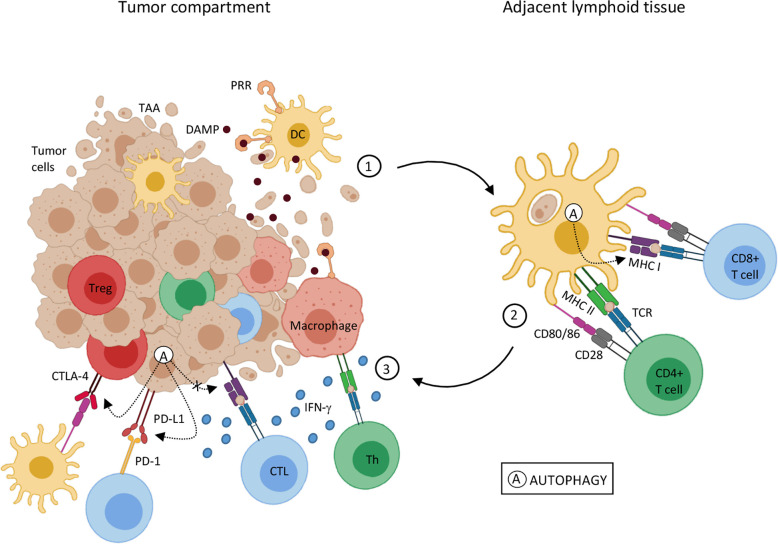
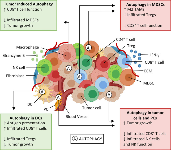
The tumor microenvironment (TME) is complex, and its composition and dynamics determine tumor fate. From tumor cells themselves, with their capacity for unlimited replication, migration, and invasion, to fibroblasts, endothelial cells, and immune cells, which can have pro and/or anti-tumor potential, interaction among these elements determines tumor progression. The understanding of molecular pathways involved in immune escape has permitted the development of cancer immunotherapies. Targeting molecules or biological processes that inhibit antitumor immune responses has allowed a significant improvement in cancer patient's prognosis. Autophagy is a cellular process required to eliminate dysfunctional proteins and organelles, maintaining cellular homeostasis. Usually a process associated with protection against cancer, autophagy associated to cancer cells has been reported in response to hypoxia, nutrient deficiency, and oxidative stress, conditions frequently observed in the TME. Recent studies have shown a paradoxical association between autophagy and tumor immune responses. Tumor cell autophagy increases the expression of inhibitory molecules, such as PD-1 and CTLA-4, which block antitumor cytotoxic responses. Moreover, it can also directly affect antitumor immune responses by, for example, degrading NK cell-derived granzyme B and protecting tumor cells. Interestingly, the activation of autophagy on dendritic cells has the opposite effects, enhancing antigen presentation, triggering CD8+ T cells cytotoxic activity, and reducing tumor growth. Therefore, this review will focus on the most recent aspects of autophagy and tumor immune environment. We describe the dual role of autophagy in modulating tumor immune responses and discuss some aspects that must be considered to improve cancer treatment.

Keywords: antitumor immunity; macroautophagy; onco immunology; tumor immune evasion; tumor microenvironment.


Conflict of interest statement

The authors declare that the research was conducted in the absence of any commercial or financial relationships that could be construed as a potential conflict of interest.

Figures

Figure 1
Figure 1
Autophagy influence on tumor-associated antigen presentation 1. Genetic alterations that cause cell transformation can also generate tumor-associated antigens (TAA) for immune recognition. Tumor cell death is an antigen source for antigen-presenting cells, such as dendritic cells (DC) and macrophages. DC activation by DAMPs and antigen processing leads to an upregulation of costimulatory molecules and MHC on DC surface, the cardinal signals for T lymphocyte activation, and migration to adjacent lymphoid tissue. 2. Mature DCs present TAA through MHC-I and MHC-II to naïve CD8+ T cells and CD4+ T cells, respectively. Antigen recognition results in T lymphocyte differentiation in effector cells (CTL e Th) and migration to the tumor site. 3. In the tumor microenvironment, upon TAA recognition through TCR interaction with MHC-I and MHC II, CTL and Th lymphocytes, respectively, trigger cytotoxic mechanisms, as interferon-gamma (IFN-γ) mediated ones. Despite that, inhibitory molecules, such as PD-1 and CTLA-4 in T cells, and PD-L1 in TME can interfere in T cell activation and function. IFN-γ stimulation can result in PD-L1 expression in both APCs and tumor cells, inhibiting T cell function. CTLA-4 expressed by regulatory T lymphocytes (Treg), through binding to co-stimulatory molecules, CD80/86, induces tolerogenic phenotype on DCs, amplifying the regulatory mechanisms in the TME. Autophagy (A) can either help or disturb the antigen presentation and T cell activation pathway. Autophagic activity on DCs seems to increase MHC-I expression, thus enhancing antigen presentation. On the other hand, autophagy activation on tumor cells may promote a reduction in MHC-I and an increase in PD-1 and CTLA-4 expression, leading to tumor progression. Sources (, , , , , –56).
Figure 2
Figure 2
Effects of autophagy on the tumor microenvironment. Tumor microenvironment (TME) is composed by cytokines (e.g. IFN-γ), extracellular matrix (ECM), and several cell types: tumor cells, fibroblasts, and immune cells, such as natural killer (NK) cells, CD8+ T and T helper (Th) lymphocytes, T regulatory (Treg) cells, myeloid-derived suppressor cells (MDSC), dendritic cells (DCs), macrophages, and pericytes (PC). Autophagy (A) is a cellular survival mechanism, which is activated in stressful conditions, can be activated in TME. Autophagy can either enhance antitumor immune responses (green boxes) or induce an immunosuppressive environment (red boxes), thus playing a dual role in cancer development and progression. Autophagy activation in DCs enhances antigen presentation and results in an enrichment of CD8+ T tumor-infiltrating lymphocytes, also decreasing infiltrated Treg and tumor cell proliferation. Similar outcomes can be seen when IFN-γ induces autophagy on tumor cells, and elevated extracellular potassium induces autophagy on CD8+ T cells. In these situations, it is possible to observe a decrease in infiltrated MDSCs and T lymphocytes expressing PD-1, thus inhibiting tumor growth. Contrarily, the autophagy activation in myeloid cells and tumor cells has the opposite effect, favoring an immunosuppressive profile of TME, leading to tumor cell proliferation, through M2 macrophage polarization, enhancing Treg infiltration and inhibitory molecules (PD-1 and CTLA-4) expression. Sources (, , , , , –67).

References

    1. Dighe AS, Richards E, Old LJ, Schreiber RD. Enhanced in vivo growth and resistance to rejection of tumor cells expressing dominant negative IFNγ receptors. Immunity (1994) 1:447–56. 10.1016/1074-7613(94)90087-6 - DOI - PubMed
    1. Shankaran V, Ikeda H, Bruce AT, White JM, Swanson PE, Old LJ, et al. IFNgamma and lympohcytes prevent primary tomour development and shape tomour immunogenicity. Nature (2001) 410:1107–11. 10.1038/35074122 - DOI - PubMed
    1. Dunn GP, Old LJ, Schreiber RD. The Three Es of Cancer Immunoediting. Annu Rev Immunol (2004) 22:329–60. 10.1146/annurev.immunol.22.012703.104803 - DOI - PubMed
    1. Schreiber RD, Old LJ, Smyth MJ. Cancer immunoediting: Integrating immunity’s roles in cancer suppression and promotion. Science (80- ) (2011) 331:1565–70. 10.1126/science.1203486 - DOI - PubMed
    1. Roma-Rodrigues C, Mendes R, Baptista PV, Fernandes AR. Targeting tumor microenvironment for cancer therapy. Int J Mol Sci (2019) 20:840. 10.3390/ijms20040840 - DOI - PMC - PubMed