Review
. 2020 Dec 5;6(6):1791-1809.
doi: 10.1016/j.bioactmat.2020.11.028. eCollection 2021 Jun.

Challenges and strategies for in situ endothelialization and long-term lumen patency of vascular grafts

Affiliations
Review

Challenges and strategies for in situ endothelialization and long-term lumen patency of vascular grafts

Yu Zhuang et al. Bioact Mater. .

Abstract

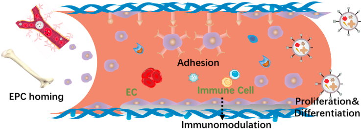
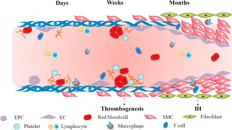
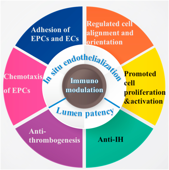
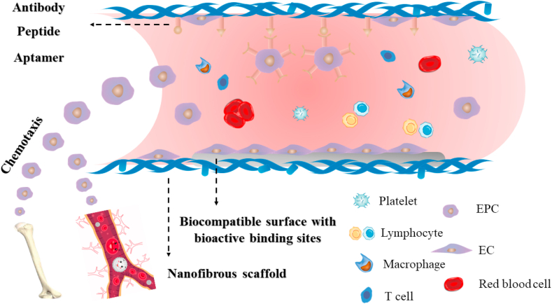
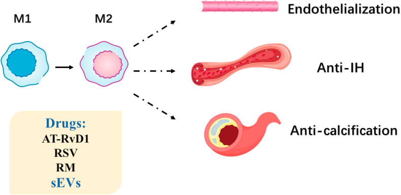
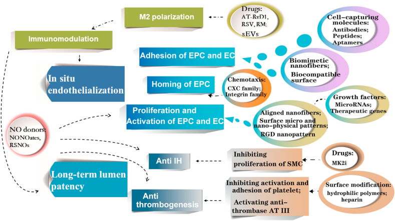
Vascular diseases are the most prevalent cause of ischemic necrosis of tissue and organ, which even result in dysfunction and death. Vascular regeneration or artificial vascular graft, as the conventional treatment modality, has received keen attentions. However, small-diameter (diameter < 4 mm) vascular grafts have a high risk of thrombosis and intimal hyperplasia (IH), which makes long-term lumen patency challengeable. Endothelial cells (ECs) form the inner endothelium layer, and are crucial for anti-coagulation and thrombogenesis. Thus, promoting in situ endothelialization in vascular graft remodeling takes top priority, which requires recruitment of endothelia progenitor cells (EPCs), migration, adhesion, proliferation and activation of EPCs and ECs. Chemotaxis aimed at ligands on EPC surface can be utilized for EPC homing, while nanofibrous structure, biocompatible surface and cell-capturing molecules on graft surface can be applied for cell adhesion. Moreover, cell orientation can be regulated by topography of scaffold, and cell bioactivity can be modulated by growth factors and therapeutic genes. Additionally, surface modification can also reduce thrombogenesis, and some drug release can inhibit IH. Considering the influence of macrophages on ECs and smooth muscle cells (SMCs), scaffolds loaded with drugs that can promote M2 polarization are alternative strategies. In conclusion, the advanced strategies for enhanced long-term lumen patency of vascular grafts are summarized in this review. Strategies for recruitment of EPCs, adhesion, proliferation and activation of EPCs and ECs, anti-thrombogenesis, anti-IH, and immunomodulation are discussed. Ideal vascular grafts with appropriate surface modification, loading and fabrication strategies are required in further studies.

Keywords: Immunomodulation; In situ endothelialization; Intimal hyperplasia; Thrombogenesis; Vascular graft.


Conflict of interest statement

The authors declare that they have no known competing financial interests or personal relationships that could have appeared to influence the work reported in this paper.

Figures

Image 1
Graphical abstract
Fig. 1
Fig. 1
The challenges after vascular graft implantation. Days and weeks after implantation, insufficient endothelialization and thrombogenesis may happen. Months after implantation, uncontrollable proliferation of SMCs may lead to IH. Inflammatory cells play a role in regulating EC and SMC behavior.
Fig. 2
Fig. 2
Schematic illustration for in situ endothelialization and lumen patency strategies.
Fig. 3
Fig. 3
Recruitment and adhesion of EPCs and ECs. Chemokines can be utilized for EPC chemotaxis. Nanofibrous structure, biocompatible surface with bioactive binding sites and specific molecules modification can be applied for EPC and EC adhesion.
Fig. 4
Fig. 4
The influence of surface topography on cell morphology and biological behavior. (A–B): Scanning electron microscopy (SEM) for inner lamellar structure of vascular graft; A: Inner lamellar structure fabricated by freeze-cast, with the lamellar 10 μm high, 200 nm thick, and the interval between lamellas was 20 μm; B: Inner non-lamellar structure fabricated by direct freeze-drying. (C): Cell behavior on graft surface; (a): Platelets adhesion. SEM figures showed that less platelets adhered on lamellar structure, and they were not activated; b: ECs elongation. ECs displayed elongated adherence along aligned surface of vascular graft and enhanced proliferation. (D): Optical figures and HE staining 3 months after implantation. Reproduced from Ref. [160], ACS NANO, ACS Publication @ 2019.
Fig. 5
Fig. 5
Bioactive molecules and therapeutic genes for enhanced in situ endothelialization. Strategies including micro/nano particle loading, nanofibers embedment or graft surface coating can be utilized to deliver therapeutic factors for promoted cell proliferation and activation. Furthermore, targeting molecules are used for more efficient gene delivery to targeted cells.
Fig. 6
Fig. 6
NO plays a crucial role in modulating endothelialization, thrombogenesis and IH. (A): The biological performance of NO. NO can be liberated by catalyzing NO donors, and play a role in vascularization, including inhibiting activation of thrombin, platelets, immune cells and proliferation of SMCs, as well as promoting proliferation and activation of ECs, relaxation and phenotype regulation of SMCs. (B–C): Fluorescence staining of ECs and SMCs 24h and 72h after in vitro culture. NO can promote EC proliferation (B) and inhibit SMC growth (C). (A) reproduced from Ref. [242], Research, CAST@ 2020. (B–C) reproduced from Ref. [249], Biomaterials, Elsevier @ 2019.
Fig. 7
Fig. 7
Macrophage performance in vascularization. Drugs or sEVs are utilized to promote the transition of M1 into M2 and regulate inflammation reactions for endothelialization enhancement, anti-IH and anti-calcification.
Fig. 8
Fig. 8
Multiple strategies for enhanced in situ endothelialization and long-term lumen patency.

References

    1. Roger V.L., Go A.S., Lloyd-Jones D.M., Adams R.J., Berry J.D., Brown T.M., Carnethon M.R., Dai S., de Simone G., Ford E.S., Fox C.S., Fullerton H.J., Gillespie C., Greenlund K.J., Hailpern S.M., Heit J.A., Ho P.M., Howard V.J., Kissela B.M., Kittner S.J., Lackland D.T., Lichtman J.H., Lisabeth L.D., Makuc D.M., Marcus G.M., Marelli A., Matchar D.B., McDermott M.M., Meigs J.B., Moy C.S., Mozaffarian D., Mussolino M.E., Nichol G., Paynter N.P., Rosamond W.D., Sorlie P.D., Stafford R.S., Turan T.N., Turner M.B., Wong N.D., Wylie-Rosett J. Heart disease and stroke statistics--2011 update: a report from the American Heart Association. Circulation. 2011;123(4):e18–e209. doi: 10.1161/CIR.0b013e3182009701. - DOI - PMC - PubMed
    1. Ouriel K. Peripheral arterial disease. Lancet. 2001;358(9289):1257–1264. doi: 10.1016/S0140-6736(01)06351-6. - DOI - PubMed
    1. Tu C., Das S., Baker A.B., Zoldan J., Suggs L.J. Nanoscale strategies: treatment for peripheral vascular disease and critical limb ischemia. ACS Nano. 2015;9(4):3436–3452. doi: 10.1021/nn507269g. - DOI - PMC - PubMed
    1. Szilagyi D.E., Smith R.F., Elliott J.P., Allen H.M. Long-term behavior of a dacron arterial substitute: clinical, roentgenologic and histologic correlations. Ann. Surg. 1965;162(3):453–477. doi: 10.1097/00000658-196509000-00015. - DOI - PMC - PubMed
    1. Zilla P., Bezuidenhout D., Human P. Prosthetic vascular grafts: wrong models, wrong questions and no healing. Biomaterials. 2007;28(34):5009–5027. doi: 10.1016/j.biomaterials.2007.07.017. - DOI - PubMed

LinkOut - more resources