Review
. 2020 Dec 18;8(Suppl 1):19.
doi: 10.1186/s40635-020-00307-1.

Pathophysiology and clinical consequences of arterial blood gases and pH after cardiac arrest

Affiliations
Review

Pathophysiology and clinical consequences of arterial blood gases and pH after cardiac arrest

Chiara Robba et al. Intensive Care Med Exp. .

Abstract

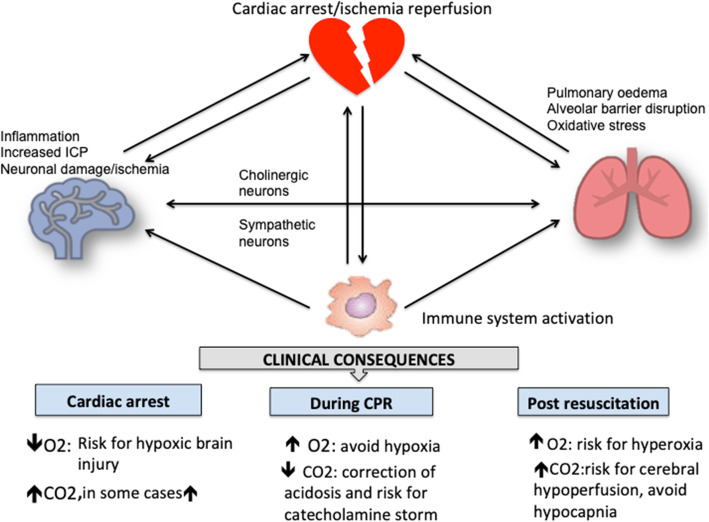
Post cardiac arrest syndrome is associated with high morbidity and mortality, which is related not only to a poor neurological outcome but also to respiratory and cardiovascular dysfunctions. The control of gas exchange, and in particular oxygenation and carbon dioxide levels, is fundamental in mechanically ventilated patients after resuscitation, as arterial blood gases derangement might have important effects on the cerebral blood flow and systemic physiology.In particular, the pathophysiological role of carbon dioxide (CO2) levels is strongly underestimated, as its alterations quickly affect also the changes of intracellular pH, and consequently influence metabolic energy and oxygen demand. Hypo/hypercapnia, as well as mechanical ventilation during and after resuscitation, can affect CO2 levels and trigger a dangerous pathophysiological vicious circle related to the relationship between pH, cellular demand, and catecholamine levels. The developing hypocapnia can nullify the beneficial effects of the hypothermia. The aim of this review was to describe the pathophysiology and clinical consequences of arterial blood gases and pH after cardiac arrest.According to our findings, the optimal ventilator strategies in post cardiac arrest patients are not fully understood, and oxygen and carbon dioxide targets should take in consideration a complex pattern of pathophysiological factors. Further studies are warranted to define the optimal settings of mechanical ventilation in patients after cardiac arrest.

Keywords: Cardiac arrest; Catecholamine; Gas exchanges; Intracellular acidosis; Ventilator targets.


Conflict of interest statement

None

Figures

Fig. 1
Fig. 1
Biological effects of pH on microglia, cardiomyocytes, and lung epithelial cells. Microglia expresses two classes of acid-base transporting proteins: acid loaders (red) and acid extruders (blue). In acidosis, acid extruder proteins are increased while in alkalosis these are downregulated. In cardiomyocytes, the regulation of the adrenergic receptors is determined by β-arrestins, which can trigger the internalization process of dephosphorization or degradation and therefore define the up- and downregulation of the internalized molecule either to recycling or degradation, respectively. In acidosis, adrenergic receptors are downregulated and less responsive to catecholamines. In the lung epithelium, reduction of lung edema clearance is associated with the endocytosis of the Na+/K+-ATPase from the plasma membrane of alveolar epithelial cells, which leads to decreased Na+/K+-ATPase activity. During acidosis, protein kinase C (PKC)-ζ phosphorylates the Na+/K+-ATPase α1-subunit, leading to endocytosis of the Na+/K+-ATPase. The activation of PKC-ζ is regulated by AMP kinase (AMPK). Acidosis culminates in the Na+/K+-ATPase endocytosis from the cell plasma membrane. In the lung epithelium, alkalosis with low carbon dioxide and hyperventilation can determine increase of mechanical power and activation of the inflammatory system
Fig. 2
Fig. 2
Effect of oxygen on microglia, cardiomyocytes, and lung epithelial cells. Microglia: hypoxic neurons have an anaerobic metabolism with increased intracellular Ca2+ intake; persistent hypoxia generates reduction of ATP and further energetic failure. Hyperoxia can disrupt microglia function by increasing free radicals. Hyperoxia increases input resistance to antioxidant and decrease membrane conductance (K+ channel) and stimulates firing of putative central CO2/H+ chemoreceptors neurons. Cardiomyocytes: effects of anoxia on cellular energetic turnover and on intra- and extracellular environments and its effect on cardiac function. In hyperoxia, O2 radical free and reduction of nitric oxide can eventually lead to coronary vasoconstriction. In the lung epithelium, hypoxia induces pulmonary vascular remodeling, with resident vascular cell activation, monocytes/fibrocytes recruitment, and persistent vasoconstriction structural remodeling. Hyperoxia exposure stimulates p53 to activate miR34a in a positive feedback loop, with consequent abnormal cell proliferation, apoptosis, impaired alveolarization, and cell death
Fig. 3
Fig. 3
Effect of carbon dioxide on microglia, cardiomyocytes, and lung epithelial cells. Microglia: hypercapnia decreases extracellular pH and intracellular pH (Phi). Increased Phi increases firing rate of CO2/H2 chemosensitive neurons, by an oxidant-induced decrease in K+ conductance. Hypocapnia with cerebral vasoconstriction and ischemic insult shifts the anaerobic metabolism and activates local and systemic inflammatory response. Cardiomyocytes: hypercapnia and acidosis reduce the sensitivity of the adrenergic receptor and expression. Hypocapnia has effect on intracellular buffers (mostly hemoglobin within red blood cells) determining release of hydrogen. Hydrogen combines with bicarbonate to form carbonic acid, which then disassociates to form water and CO2, thus replenishing the depleted PaCO2. Lung epithelium: hypercapnia inhibits proliferation of alveolar epithelial cells due to mitochondrial dysfunction resulting from hypercapnia-induced miR-183 which downregulates the TCA cycle enzyme isocitrate dehydrogenase-2 (IDH). Hypercapnic acidosis impairs alveolar epithelial cell migration by the NF-kB dependent mechanism. Hypercapnia inhibits mRNA and protein expression of IL-6 and TNF and decreases phagocytosis in macrophages. Hypocapnia and hyperventilation can determine increase of mechanical power and activation of the inflammatory system. CO2, carbon dioxide; PaCO2, partial pressure of carbon dioxide; TCA, tricarboxylic acid cycle; NF-kB, nuclear factor kappa-light-chain-enhancer of activated B cells; IDH, enzyme isocitrate dehydrogenase-2; TNF, tumor necrosis factor; IL, interleukin; mRNA, ribonucleic acid
Fig. 4
Fig. 4
The pathophysiologic vicious circle after cardiac arrest: hyperventilation and hypocapnia can quickly cause intracellular and extracellular pH increase; this increases metabolic energy and O2 demand, while ATP production is reduced in cardiac arrest for the activation of the anaerobic pathway; this determinates a sympathetic-like activation aimed to compensate the increased energy demand. Also, cardiac arrest per se' and pH derangements strongly influence intracellular function, thus increasing metabolic expenditure, and oxygen consumption. This occurs in the contest of cardiac injury related to the cardiac arrest as well as to pH changes which can further impair cardiac function; impaired myocardium function results in impaired diastolic function with further hyperventilation and hypocapnia. O2, oxygen; ATP, adenosine triphosphate
Fig. 5
Fig. 5
Systemic effect of cardiac arrest and clinical implications in the ventilator targets to achieve. Catecholamine release, with impaired mitochondrial metabolism, can activate immunology response. Neuronal and immune responses are rapidly activated during cardiac arrest and trigger a pathway of systemic neuroendocrine and immune responses which have important systemic consequences. Global cerebral ischemia and immunological disturbances induce microgliosis, damage of blood-brain barrier, and cerebrovascular system. Endothelial dysfunction and blood brain barrier permeability are crucial in above pathological processes. The brain-lung or brain-heart-lung couplings and their disturbances may be considered as modulator of post ischemic injury, peripheral inflammation, and multiorgan dysfunction, observed in post cardiac arrest patients. Post ischemic induction of necrosis, apoptosis, and systemic inflammation predisposes to neuronal damage and poor recovery. Pulmonary complications following cardiac arrest, through cerebral ischemia or immunological activation, can result in neurogenic pulmonary edema, ARDS, or pneumonia. The clinical consequences of these pathophysiological pathways are different according to three different phases: cardiac arrest, cardiopulmonary resuscitation, and post resuscitation. ARDS, acute respiratory distress syndrome

References

    1. Relman AS. Metabolic consequences of acid-base disorders. Kidney Int. 1972;1:347–359. doi: 10.1038/ki.1972.46. - DOI - PubMed
    1. Kimmoun A, Novy E, Auchet T, et al. Hemodynamic consequences of severe lactic acidosis in shock states: from bench to bedside. Crit Care. 2015;19:175. doi: 10.1186/s13054-015-0896-7. - DOI - PMC - PubMed
    1. Phypers B, Pierce JT. Lactate physiology in health and disease. Contin Educ Anaesth Crit Care Pain. 2006;6:128–132. doi: 10.1093/bjaceaccp/mkl018. - DOI
    1. Levy B. Lactate and shock state: the metabolic view. Curr Opin Crit Care. 2006;12:315–321. doi: 10.1097/01.ccx.0000235208.77450.15. - DOI - PubMed
    1. Wutrich Y, Barraud D, Conrad M, et al. Early increase in arterial lactate concentration under epinephrine infusion is associated with a better prognosis during shock. Shock. 2010;34:4–9. doi: 10.1097/SHK.0b013e3181ce2d23. - DOI - PubMed

LinkOut - more resources