Review
. 2021;8(2):185-207.
doi: 10.3233/JND-200490.

Current View of Diagnosing Small Fiber Neuropathy

Affiliations
Review

Current View of Diagnosing Small Fiber Neuropathy

Lisette R M Raasing et al. J Neuromuscul Dis. 2021.

Abstract

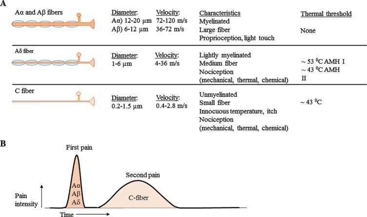
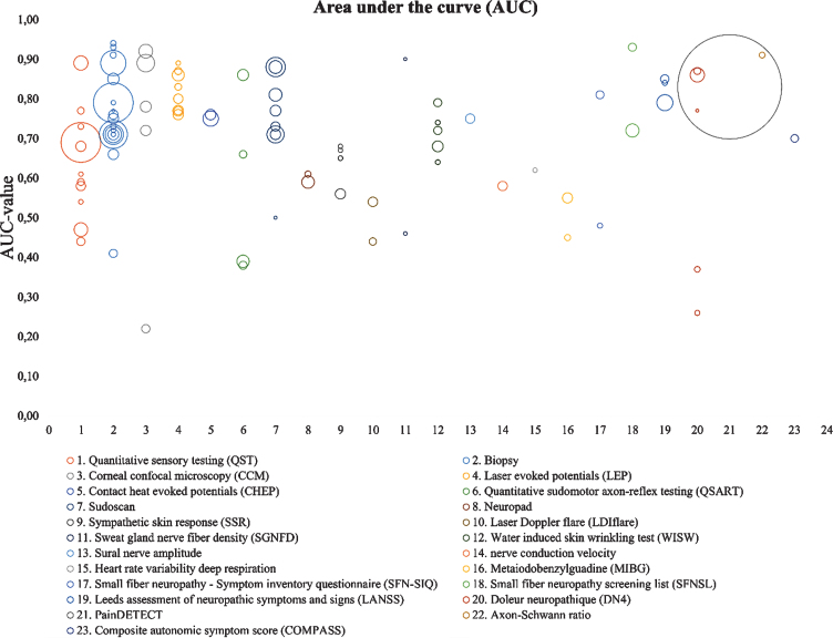
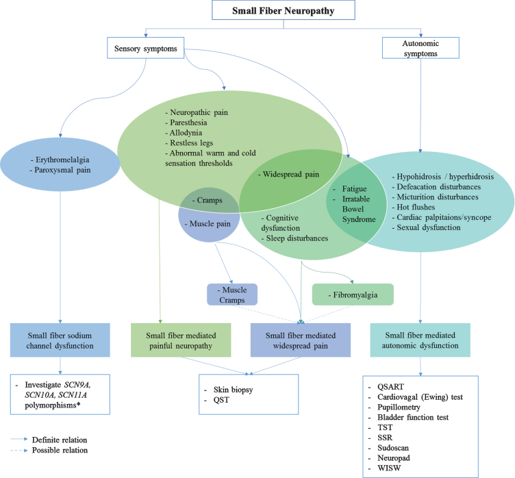
Small fiber neuropathy (SFN) is a disorder of the small myelinated Aδ-fibers and unmyelinated C-fibers [5, 6]. SFN might affect small sensory fibers, autonomic fibers or both, resulting in sensory changes, autonomic dysfunction or combined symptoms [7]. As a consequence, the symptoms are potentially numerous and have a large impact on quality of life [8]. Since diagnostic methods for SFN are numerous and its pathophysiology complex, this extensive review focusses on categorizing all aspects of SFN as disease and its diagnosis. In this review, sensitivity in combination with specificity of different diagnostic methods are described using the areas under the curve. In the end, a diagnostic work-flow is suggested based on different phenotypes of SFN.

Keywords: Autonomic dysfunction; diagnostic accuracy; nerve fiber density; small fiber neuropathy.


Conflict of interest statement

The authors declare no competing financial interests

Figures

Fig. 1
Fig. 1
A) Overview of nerve fiber sizes, conduction velocities and other characteristics. Aα and Aβ fibers are large and myelinated nerve fibers, Aδ nerve fibers are small myelinated nerve fibers and C-fibers are small unmyelinated nerve fibers. B) Corresponding pain response. Large nerve fibers show a fast response with high amplitude. The smaller the nerve fiber, the lower the amplitude and the slower conduction velocities.
Fig. 2
Fig. 2
Complete overview of the nervous system, showing anatomical differences between the somatic and autonomic system and differences in nerve anatomy and use of neurotransmitters. In addition, all diagnostic methods are presented at their corresponding measuring area. Different font styles are used to discriminate between methods based on NFD (bold), small nerve fiber function (italic) and imaging (normal). Moreover, for some functional tests, an additional mark is established to discriminate between tests based on thermal and/or mechanical nociceptors. Abbreviations: EPs, evoked potentials; fMRI, functional magnetic resonance imaging; TST, thermoregulatory sweat testing; US, ultrasound; CCM, corneal confocal microscopy; IENFD, intra-epidermal nerve fiber density; QST, quantitative sensory testing; TTT, temperature threshold testing; HR, heartrate; EMG, electromyography; MIBG, 123I-meta-iodobenzylguadine; QPART, quantitative pilomotor axon-reflex test; BP, blood pressure; SGNFD, sweat gland nerve fiber density; LDIflare, laser Doppler imaging flare; QSART, quantitative sensory axon reflex test; QDIRT, quantitative direct and indirect reflex test; SSR, sympathetic skin response.
Fig. 3
Fig. 3
Inclusion results of systematic literature search. Articles excluded with a wrong study design, included review articles, animal models or case reports. If review articles did publish data about sensitivity (sens) and/or specificity (spec), the original papers were looked up. As a result, 29 extra articles were included. 19 articles did publish the test results from all participants, which made it possible to calculate sens & spec. 57 articles did calculate sens & spec based on QST and/or IENFD.
Fig. 4
Fig. 4
Overview of neuropathy questionnaires, divided into screening questionnaires, assessment questionnaires, specific small fiber questionnaires and pain intensity questionnaires.
Fig. 5
Fig. 5
AUC overview of several diagnostic methods. It determines the diagnostic accuracy of each method. The size of each bubble, represents the study size. Unmyelinated axon-Schwann ration is calculated based on biopsies and not further reviewed in this article.
Fig. 6
Fig. 6
Roadmap; from symptoms to phenotype, from phenotype to diagnostic method. * Many variants are probably nonpathogenic or variant of undetermined significance (VUS). Abbreviations: QSART, quantitative sensory axon reflex test; TST, thermoregulatory sweat test; SSR, sympathetic skin response; WISW, water induced skin wrinkling.

Similar articles

Cited by

References

    1. Eijkenboom I, Sopacua M, Hoeijmakers JGJ, et al.. Yield of peripheral sodium channels gene screening in pure small fibre neuropathy. J Neurol Neurosurg Psychiatry. 2019;90(3):342–52. doi:10.1136/jnnp-2018-319042 - DOI - PubMed
    1. Fabry V, Gerdelat A, Acket B, et al.. Which Method for Diagnosing Small Fiber Neuropathy? Front Neurol. 2020;11(May):1–9. doi:10.3389/fneur.2020.00342 - DOI - PMC - PubMed
    1. Devigili G, Cazzato D, Lauria G. Clinical diagnosis and management of small fiber neuropathy: An update on best practice. Expert Rev Neurother. 2020;00(00):1–14. doi:10.1080/14737175.2020.1794825 - DOI - PubMed
    1. Devigili G, Rinaldo S, Lombardi R, et al.. Diagnostic criteria for small fibre neuropathy in clinical practice and research. Brain. 2019;142(12):3728–36. doi:10.1093/brain/awz333 - DOI - PMC - PubMed
    1. Hovaguimian A, Gibbons CH. Diagnosis and Treatment of Pain in Small-Fiber Neuropathy. Vol 15.; 2011. doi:10.1007/s11916-011-0181-7 - DOI - PMC - PubMed