. 2021 Aug;15(8):2106-2119.
doi: 10.1002/1878-0261.12888. Epub 2021 Jan 4.

Pharmacologic inhibition of Akt in combination with chemotherapeutic agents effectively induces apoptosis in ovarian and endometrial cancer cell lines

Affiliations

Pharmacologic inhibition of Akt in combination with chemotherapeutic agents effectively induces apoptosis in ovarian and endometrial cancer cell lines

François Fabi et al. Mol Oncol. 2021 Aug.

Abstract

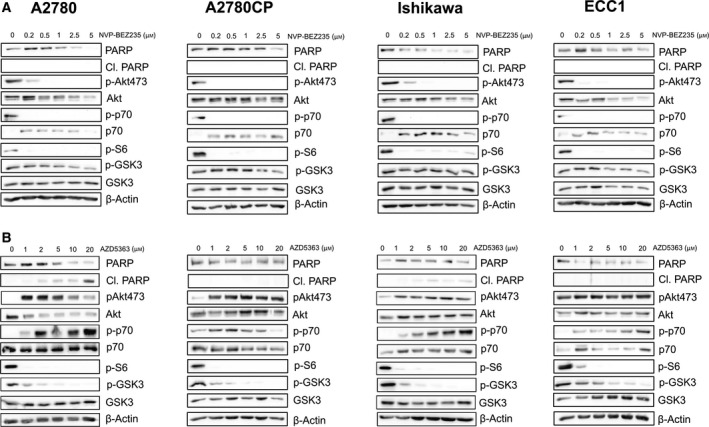
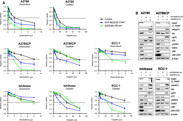
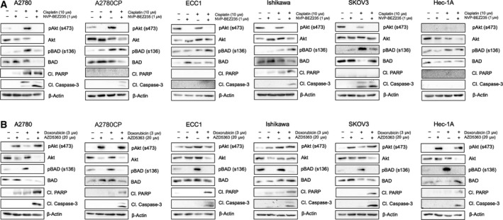
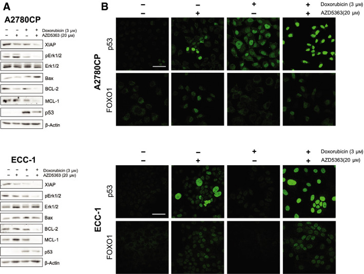
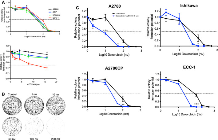
The PI3K/Akt signaling pathway, the most frequently altered signaling system in human cancer, is a crucial inducer of dysregulated proliferation and neoplastic processes; however, few therapeutic strategies using PI3K/Akt inhibitors singly have been shown to be effective. The purpose of this paper was to underline the potential benefit of pharmacological modulation of the PI3K/Akt pathway when combined with specific chemotherapeutic regimens. We have studied the ability of NVP-BEZ235 (PI3K/mTOR inhibitor) and AZD5363 (Akt inhibitor) in the sensitization of cancer cells to cisplatin and doxorubicin. Our results show that NVP-BEZ235 sensitizes cells preferentially to cisplatin while AZD5363 sensitizes cells to doxorubicin. At equal concentrations (5 μm), both inhibitors reduce ribosomal protein S6 phosphorylation, but AZD5363 is more effective in reducing GSK3β phosphorylation as well as S6 phosphorylation. Additionally, AZD5363 is capable of inducing FOXO1 and p53 nuclear localization and reduces BAD phosphorylation, which is generally increased by cisplatin and doxorubicin. Finally, the combination of AZD5363 and doxorubicin induces apoptosis in cells and robustly reduces cell ability to clonally replicate, which underlines a potential cooperative effect of the studied compounds.

Keywords: AZD5363; capivasertib; chemoresistance; doxorubicin; endometrial cancer; ovarian cancer.


Conflict of interest statement

The authors declare no conflict of interest.

Figures

Fig. 1
Fig. 1
NVP‐BEZ235 and AZD5363 modulate signaling pathways in a dose‐dependent manner. (A) Cell lines were treated with increasing concentration of NVP‐BEZ235 (0–5 μm). Western blot was performed using relevant antibodies, and β‐actin was used as a loading control. Results shown are representative of three independent experiments. (B) Cell lines were treated with increasing concentration of AZD5363 (0–20 μm). Western blot was performed using relevant antibodies, and β‐actin was used as a loading control. Results shown are representative of three independent experiments. [Colour figure can be viewed at wileyonlinelibrary.com]
Fig. 2
Fig. 2
Combination of AZD5363 or NVP‐BEZ235 sensitizes cells to cisplatin and doxorubicin. (A) Cell lines were treated with increasing concentration of cisplatin (0–80 μm) or doxorubicin (0–8 μm) in the presence or absence of either AZD5363 (20 μm) or NVP‐BEZ‐235 (1 μm) for 24 h. MTT assays were then used to determine changes in cell viability. (B) Cell lines were treated with equal concentration of NVP‐BEZ235 or AZD5363 (5 μm). Western blot was performed using relevant antibodies, and β‐actin was used as a loading control. One‐way ANOVA followed by Tukey's post hoc test was performed. All data are means ± SEM of three independent experiments. *P < 0.05. [Colour figure can be viewed at wileyonlinelibrary.com]
Fig. 3
Fig. 3
Combination of AZD5363 with doxorubicin induces apoptosis in gynecological cancer cell lines. (A) Cell lines were treated with either cisplatin (10 μm), NVP‐BEZ‐235 (1 μm), or a combination of both for 24 h. Western blot was performed using relevant antibodies, and β‐actin was used as a loading control. (B) Cell lines were treated with either doxorubicin (3 μm), AZD5363 (20 μm), or a combination of both for 24 h. Western blot was performed using relevant antibodies, and β‐actin was used as a loading control. Results shown are representative of three independent experiments. [Colour figure can be viewed at wileyonlinelibrary.com]
Fig. 4
Fig. 4
Combination of AZD5363 with doxorubicin regulates key pro‐apoptotic signaling pathways. (A) Cell lines were treated with either doxorubicin (3 μm), AZD5363 (20 μm), or a combination of both for 24 h. Western blot was performed using relevant antibodies, and β‐actin was used as a loading control. Results shown are representative of three independent experiments. (B) Immunofluorescence experiments were conducted in order to determine the effect of the previous treatments on p53 and FOXO1 subcellular localization. Scale bar = 45 μm. [Colour figure can be viewed at wileyonlinelibrary.com]
Fig. 5
Fig. 5
AZD5363 potentiates the ability of doxorubicin to reduce cell proliferation and clonal replication. (A) Cell lines were treated with increasing concentrations of AZD5363 (0–16 μm) or doxorubicin (0–500 nm). (B) Representative clonogenic assay; in this instance, all cells were pretreated with AZD5363 (5 μm) and with increasing concentrations of doxorubicin (0–200 nm). Densitometric analysis was conducted using colonyarea software. (C) Cell lines were treated with increasing concentrations of doxorubicin (0–200 nm) in the presence of AZD5363 (5 μm). Densitometric analysis was conducted using colonyarea software. Two‐way ANOVA followed by Tukey's post hoc test was performed. All data are means ± SEM of three independent experiments. *P < 0.05, **P < 0.01, ***P < 0.001. Corresponding calculated IC50 can be found in Table 1. [Colour figure can be viewed at wileyonlinelibrary.com]

Similar articles

Cited by

References

    1. Bailey MH, Tokheim C, Porta‐Pardo E, Sengupta S, Bertrand D, Weerasinghe A, Colaprico A, Wendl MC, Kim J, Reardon B et al, (2018) Comprehensive Characterization of Cancer Driver Genes and Mutations. Cell 173(2), 371–385 e18. - PMC - PubMed
    1. Manning BD & Toker A (2017) AKT/PKB Signaling: Navigating the Network. Cell 169(3), 381–405. - PMC - PubMed
    1. Tsherniak A, Vazquez F, Montgomery PG, Weir BA, Kryukov G, Cowley GS, Gill S, Harrington WF, Pantel S, Krill‐Burger JM et al, (2017) Cell 170(3), 564–576 e16. - PMC - PubMed
    1. Gagnon V, Mathieu I, Sexton E, Leblanc K & Asselin E (2004) AKT involvement in cisplatin chemoresistance of human uterine cancer cells. Gynecol Oncol 94(3), 785–795. - PubMed
    1. Kim D, Dan HC, Park S, Yang L, Liu Q, Kaneko S, Ning J, He L, Yang H, Sun M et al, (2005) AKT/PKB signaling mechanisms in cancer and chemoresistance. Front Biosci 10, 975–987. - PubMed

Publication types

MeSH terms

Substances