Multicenter Study
. 2021 Mar;5(3):167-177.
doi: 10.1016/S2352-4642(20)30362-X. Epub 2020 Dec 16.

Neuroimaging manifestations in children with SARS-CoV-2 infection: a multinational, multicentre collaborative study

Collaborators, Affiliations
Multicenter Study

Neuroimaging manifestations in children with SARS-CoV-2 infection: a multinational, multicentre collaborative study

Camilla E Lindan et al. Lancet Child Adolesc Health. 2021 Mar.

Abstract

Background: The CNS manifestations of COVID-19 in children have primarily been described in case reports, which limit the ability to appreciate the full spectrum of the disease in paediatric patients. We aimed to identify enough cases that could be evaluated in aggregate to better understand the neuroimaging manifestations of COVID-19 in the paediatric population.

Methods: An international call for cases of children with encephalopathy related to severe acute respiratory syndrome coronavirus 2 (SARS-CoV-2) infection and abnormal neuroimaging findings was made. Clinical history and associated plasma and cerebrospinal fluid data were requested. These data were reviewed by a central neuroradiology panel, a child neurologist, and a paediatric infectious diseases expert. The children were categorised on the basis of their time of probable exposure to SARS-CoV-2. In addition, cases were excluded when a direct link to SARS-CoV-2 infection could not be established or an established alternate diagnostic cause could be hypothesised. The accepted referral centre imaging data, from ten countries, were remotely reviewed by a central panel of five paediatric neuroradiologists and a consensus opinion obtained on the imaging findings.

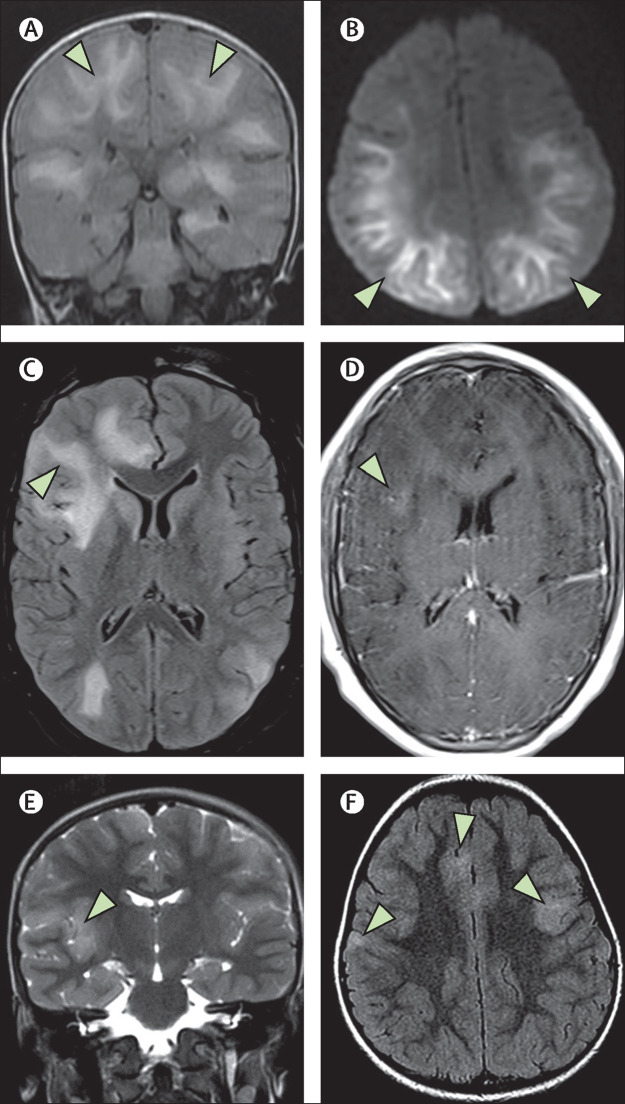
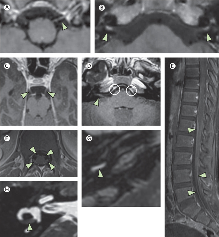
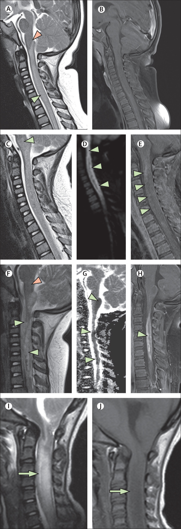
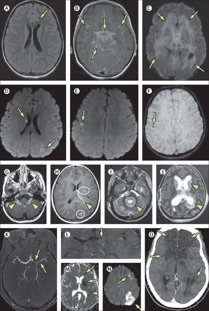
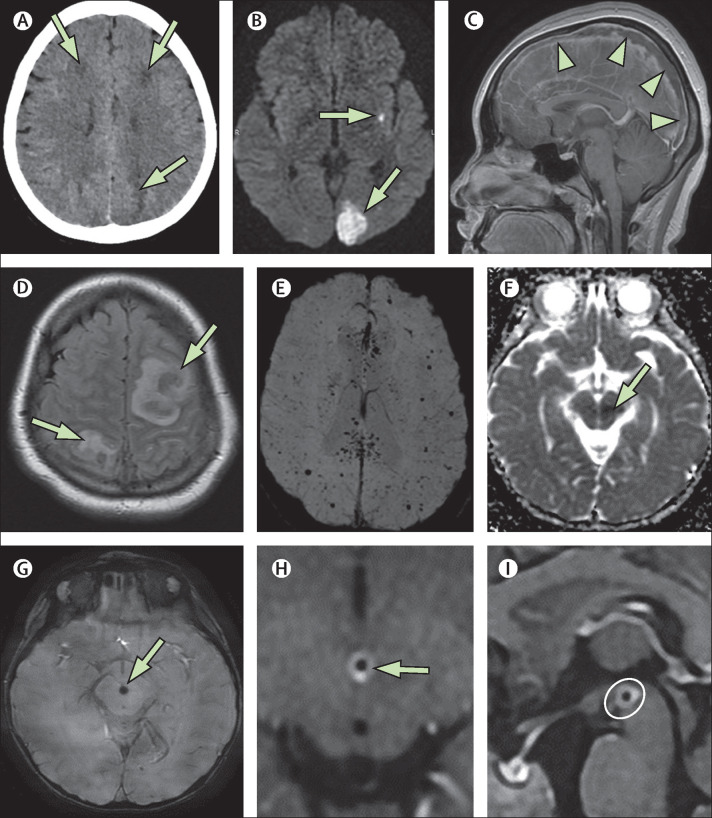
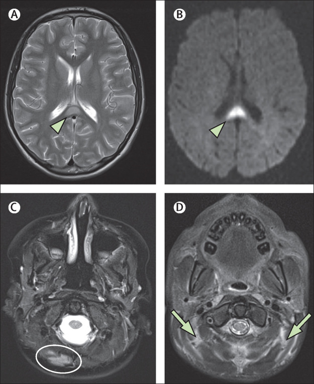
Findings: 38 children with neurological disease related to SARS-CoV-2 infection were identified from France (n=13), the UK (n=8), the USA (n=5), Brazil (n=4), Argentina (n=4), India (n=2), Peru (n=1), and Saudi Arabia (n=1). Recurring patterns of disease were identified, with neuroimaging abnormalities ranging from mild to severe. The most common imaging patterns were postinfectious immune-mediated acute disseminated encephalomyelitis-like changes of the brain (16 patients), myelitis (eight patients), and neural enhancement (13 patients). Cranial nerve enhancement could occur in the absence of corresponding neurological symptoms. Splenial lesions (seven patients) and myositis (four patients) were predominantly observed in children with multisystem inflammatory syndrome. Cerebrovascular complications in children were less common than in adults. Significant pre-existing conditions were absent and most children had favourable outcomes. However, fatal atypical CNS co-infections developed in four previously healthy children infected with SARS-CoV-2.

Interpretation: Acute-phase and delayed-phase SARS-CoV-2-related CNS abnormalities are seen in children. Recurring patterns of disease and atypical neuroimaging manifestations can be found and should be recognised being as potentially due to SARS-CoV-2 infection as an underlying aetiological factor. Studies of paediatric specific cohorts are needed to better understand the effects of SARS-CoV-2 infection on the CNS at presentation and on long-term follow-up in children.

Funding: American Society of Pediatric Neuroradiology, University of Manchester (Manchester, UK). VIDEO ABSTRACT.


Figures

Figure 1
Figure 1
ADEM-like brain changes (A, B) A 1-year-old boy (case 2) with acute COVID-19 showed confluent areas of high signal in the subcortical white matter on coronal FLAIR imaging (A; arrows), and reduced diffusion on DWI trace (B; arrows). (C, D) A 13-year-old boy (case 4) showed similar changes on FLAIR imaging with associated mass effect in the right frontal lobe (C; arrow). This area showed some subtle enhancement on postcontrast T1-weighted imaging (D; arrow).(E, F) In a 4-year-old boy (case 38) with an indeterminate timepoint of exposure to SARS-CoV-2, ADEM-like changes were seen on coronal T2-weighted images (E; arrow) and axial FLAIR images (F; arrows). This child was positive for antibodies to myelin oligodendrocyte glycoprotein. ADEM=acute disseminated encephalomyelitis. DWI=diffusion-weighted imaging. FLAIR=fluid-attenuated inversion recovery.
Figure 2
Figure 2
Neuritis (A, B) A 5-year-old boy (case 6) with acute COVID-19 presented with acute facial paralysis in conjunction with respiratory failure. He had marked enhancement and thickening of multiple cranial nerves, for example the 12th nerve on the left (A; arrowhead) and the seventh nerves bilaterally (B; arrowheads). (C–F) A 9-year-old boy (case 7), also with acute COVID-19, showed similar cranial nerve enhancement of his third nerves (C; arrowheads) as well as his seventh and eighth nerves (D; arrowhead [shown on patient's right side]) and his sixth nerves bilaterally (D; circles). This child also had enhancement of the cauda equina (E; arrowheads) as well as his cervical spine nerve roots (F; arrowheads). (G, H) A 13-year-old boy (case 33) with labyrinthitis with enhancement of the basal turn of the cochlea (G; arrowhead) and partial obliteration of his horizontal semicircular canal (H; arrowhead). All panels show T1 postcontrast images except for panel H (fast-spin echo T2 image).
Figure 3
Figure 3
Acute necrotising myelitis (A–H) A 3-year-old girl (case 5), who was living in a household with multiple family members who had COVID-19, presented with acute SARS-CoV-2 infection with positive PCR result. Symptoms included acute respiratory failure, confusion, limb weakness, and vomiting. Initial T2-weighted imaging (A) showed central cervical cord signal abnormality (green arrowhead) extending up to the obex (pink arrowhead) but sparing the medulla. No enhancement was seen in the cervical and thoracic cord on the initial T1-weighted postcontrast imaging (B). 4 days later, more extensive myelitis was seen with new involvement of the medulla on T2 imaging (C; arrowhead), new reduced diffusion seen on diffusion trace images (D; arrowheads), and progressive enhancement seen on T1 postcontrast imaging (E; arrowheads). 3·5 weeks later, marked cord atrophy and necrosis were seen on sagittal T2 imaging (F; green arrowheads) with resolution of the medullary signal change (pink arrowhead). Persistent and varied areas of reduced diffusion were seen on sagittal apparent diffusion coefficient maps (G; arrowheads) in addition to enhancement on T1 postcontrast sagittal imaging (H; arrowhead), suggesting ongoing active disease. (I, J) For comparison, a second case of severe myelitis in a 5-year-old girl (case 8) with acute COVID-19 is shown. Sagittal T2-weighted (I) and sagittal T1-weighted (J) images show profound cord swelling (arrowheads). This child (case 8) died with biopsy-proven tuberculous granulomata and electron microscopic evidence of SARS-CoV-2 viral inclusions in the brain. SARS-CoV-2=severe acute respiratory syndrome coronavirus 2.
Figure 4
Figure 4
Fatal co-infections (A–C) 5-year-old girl (case 8) presented with fever, headache, and seizures. Initial MRI showed an acute small left frontal infarct on axial FLAIR imaging (A; arrow). 5 days later she had extensive leptomeningeal enhancement in the basilar cisterns and perisylvian regions on postcontrast T1-weighted imaging (B; arrows). Findings progressed and, 3 weeks after presentation, markedly reduced diffusion on diffusion trace imaging (C; arrows) and oedema of the brain parenchyma were observed. The patient's brain biopsy was positive for SARS-CoV-2 viral inclusions on electron microscopy and positive for tuberculosis granulomata despite no tuberculosis contact. (D–F) A 5-year-old girl (case 9) presented with encephalopathy and acute respiratory distress and became septic with meticillin-resistant Staphylococcus aureus and varicella zoster virus infections, both of which were culture positive in the blood and CSF. She had multiple small foci of reduced diffusion on axial diffusion trace imaging, in keeping with microinfarcts (D, E; arrows), some of which had associated microhaemorrhage (F; arrow). This patient died 15 days after symptom onset. (G–J) A 6-year-old boy (case 10) with no previous comorbidities presented with scattered T2 hyperintensities in the supratentorial white matter. He also had marked choroid plexitis (G; arrowheads) and foci of ring enhancement on T1 postcontrast imaging, in keeping with small abscesses (H; circles). There was minimal ependymal enhancement (H; arrowhead). Reduced diffusion was noted in the affected choroid and abscesses (not shown). 2 weeks later, axial T2-weighted imaging showed extensive and rapid progression involving multiple supratentorial and infratentorial compartments with ependymal invasion around the fourth ventricle (I; arrowhead), the basal ganglia, and the lateral ventricles (J; arrowhead). These areas also showed enhancement and reduced diffusion (not shown). Mycobacterium tuberculosis infection was confirmed at open biopsy. (K–O) A 16-year-old boy (case 11) presented with encephalopathy, fever, sinusitis, and meningismus, and Fusobacterium necrophorum and Streptococcus constellatus co-infections in the blood and CSF. At presentation he had multivessel stenoses on his magnetic resonance angiogram, most markedly involving the anterior and middle cerebral arteries (K; arrows), and there was associated vessel wall enhancement on postcontrast arterial wall imaging (L; arrow) and multiple vascular territory infarcts on the apparent diffusion coefficient map and diffusion-weighted trace image (M, N; arrows), features all in keeping with multivessel vasculitis. This condition progressed to multiple regional infarctions as shown on CT at day 5 (O; arrows). FLAIR=fluid-attenuated inversion recovery.
Figure 5
Figure 5
Vasculitic and thrombotic findings (A, B) A 15-year-old girl (case 12) presented 27 weeks pregnant with fever, seizures, and hypertension, and COVID-19 pneumonia. Her CT at presentation (A) showed low-density areas in multiple locations (arrows). MRI 7 days later (B) showed small focal infarcts and a larger left occipital infarct (arrows) on diffusion trace imaging, findings compatible with unusually severe posterior reversible encephalopathy syndrome. (C, D) A 15-year-old girl (case 20) with subacute COVID-19 and no classical respiratory symptoms presented with fever, confusion, and headache. Complete occlusive thrombosis of the superior sagittal sinus was shown by the large filling defect in the postcontrast sagittal T1-weighted image (C; arrowheads), with resultant bilateral haemorrhagic venous infarcts on axial FLAIR images (D; arrows). (E) A 15-year-old girl (case 27) with multisystem inflammatory syndrome in children who also developed multiple microthrombi, as shown on SWI. The microthrombi were relatively clinically silent and showed partial resolution at 3 weeks with full clinical resolution of symptoms at 3 months after presentation. (F–I) A 2-year-old girl (case 32, indeterminate category) presented with fever and pharyngeal pain with an acute left midbrain infarction (arrow) shown on the apparent diffusion coefficient map (F; arrow). She had a thrombus in the feeding anterior perforator vessel on SWI (G; arrow) and marked associated vessel wall enhancement on postcontrast T1 arterial wall imaging (H, arrow; I, circle). FLAIR=fluid-attenuated inversion recovery. SWI=susceptibility-weighted imaging.
Figure 6
Figure 6
Myositis and splenial lesions (A) Axial T2-weighted image in a 14-year-old boy (case 26) with the classic appearance of a splenial lesion (arrowhead) in post-COVID-19 MIS-C. This lesion showed reduced diffusion at presentation (arrowhead), as shown by the diffusion trace image in the same patient (B). Four patients with MIS-C were also noted to have myositis, which could be focal (C; oval), as seen in an 8-year-old boy (case 28), or diffuse (D; arrows), as seen in a 9-year-old boy (case 23) on axial T2-weighted fat-saturated images. MIS-C=multisystem inflammatory syndrome in children.

Similar articles

Cited by

References

    1. Zhu N, Zhang D, Wang W. A novel coronavirus from patients with pneumonia in China, 2019. N Engl J Med. 2020;382:727–733. - PMC - PubMed
    1. Parri N, Lenge M, Buonsenso D. Children with COVID-19 in pediatric emergency departments in Italy. N Engl J Med. 2020;383:187–190. - PMC - PubMed
    1. Götzinger F, Santiago-García B, Noguera-Julián A. COVID-19 in children and adolescents in Europe: a multinational, multicentre cohort study. Lancet Child Adolesc Health. 2020;4:653–661. - PMC - PubMed
    1. Niazkar HR, Zibaee B, Nasimi A, Bahri N. The neurological manifestations of COVID-19: a review article. Neurol Sci. 2020;41:1667–1671. - PMC - PubMed
    1. Yaghi S, Ishida K, Torres J. SARS-CoV-2 and stroke in a New York healthcare system. Stroke. 2020;51:2002–2011. - PMC - PubMed

Publication types

MeSH terms

Supplementary concepts