. 2021 Mar:144:115819.
doi: 10.1016/j.bone.2020.115819. Epub 2020 Dec 16.

GATA4 regulates mesenchymal stem cells via direct transcriptional regulation of the WNT signalosome

Affiliations

GATA4 regulates mesenchymal stem cells via direct transcriptional regulation of the WNT signalosome

Aysha B Khalid et al. Bone. 2021 Mar.

Abstract

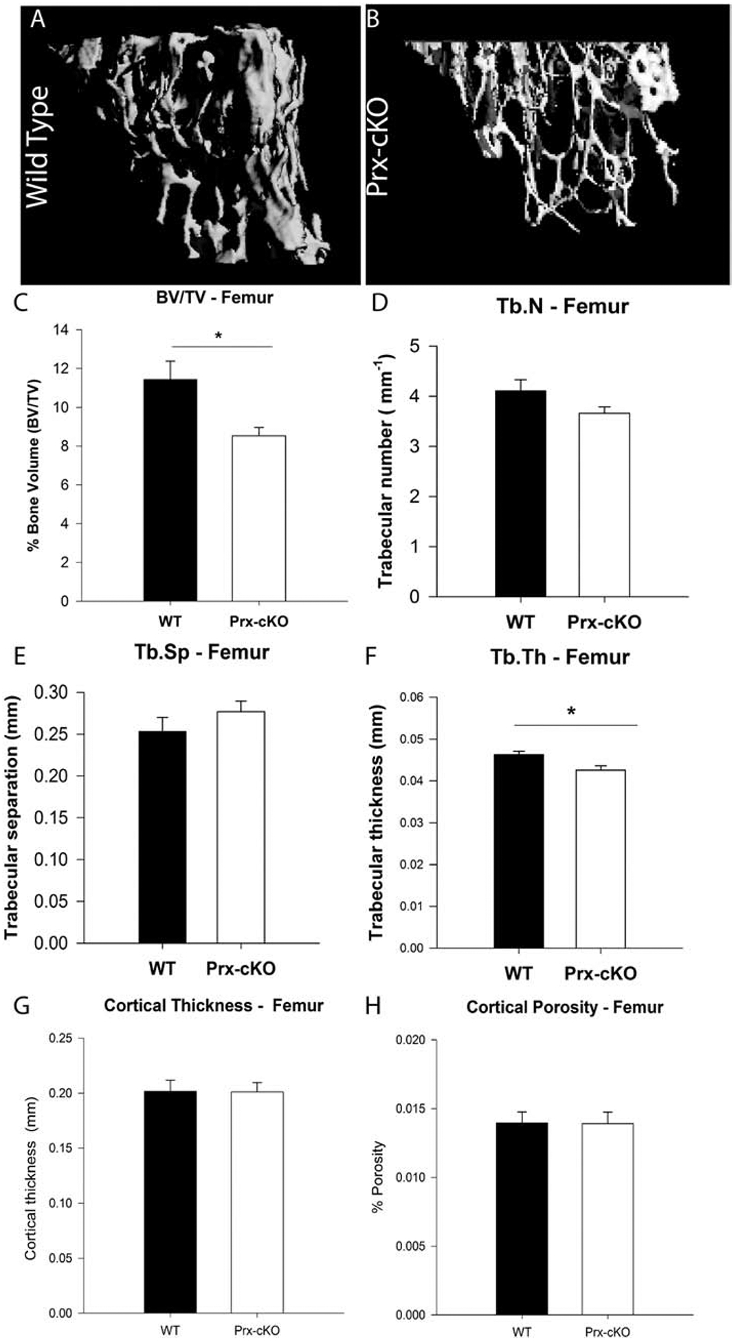
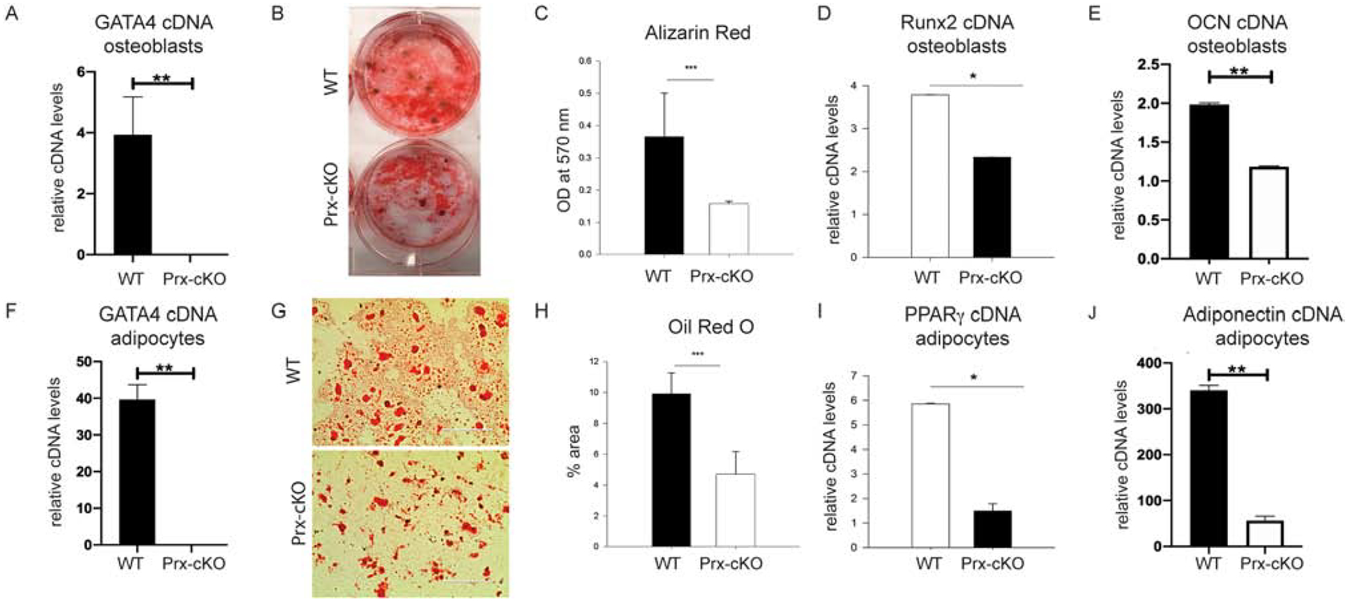
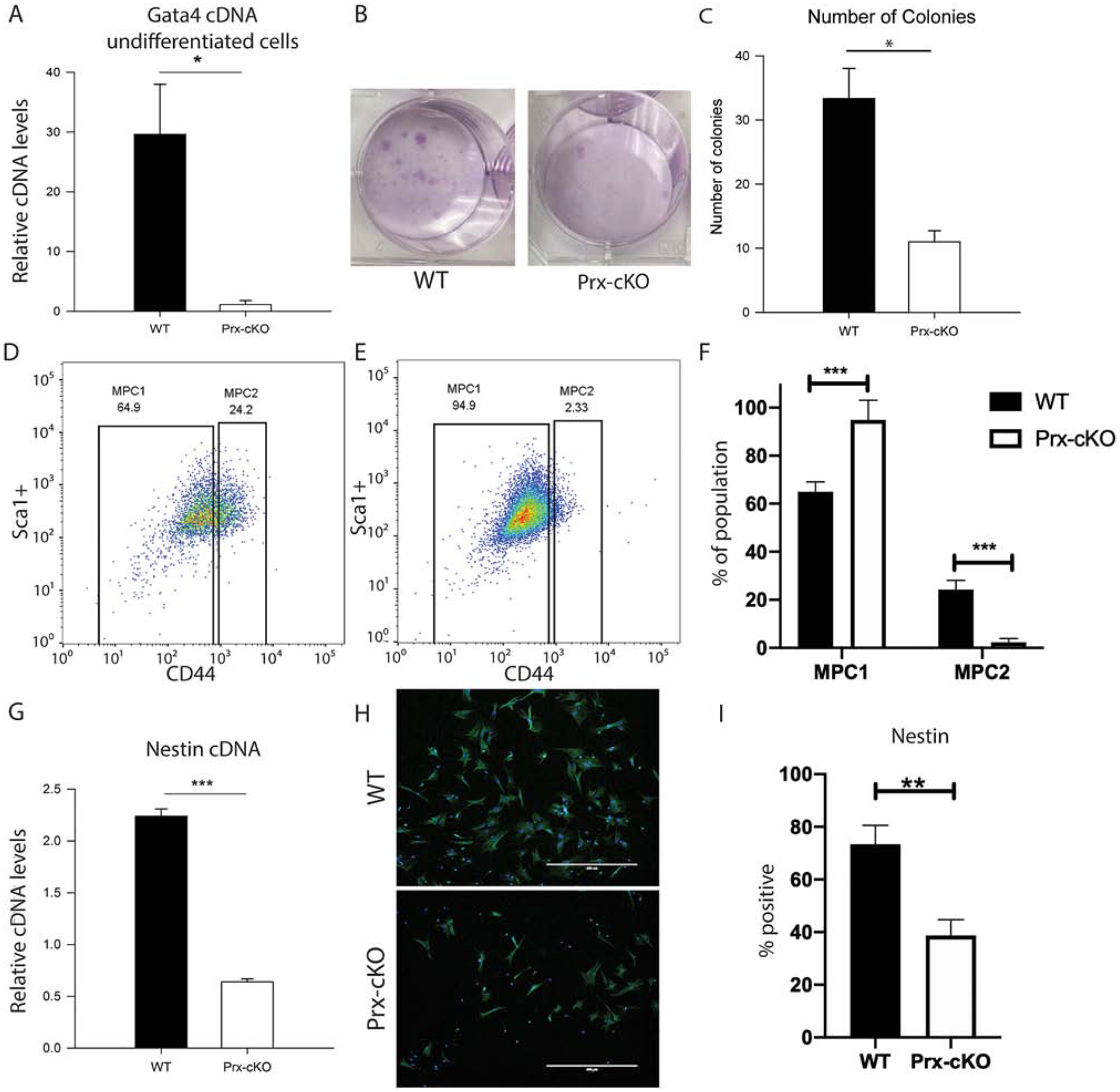
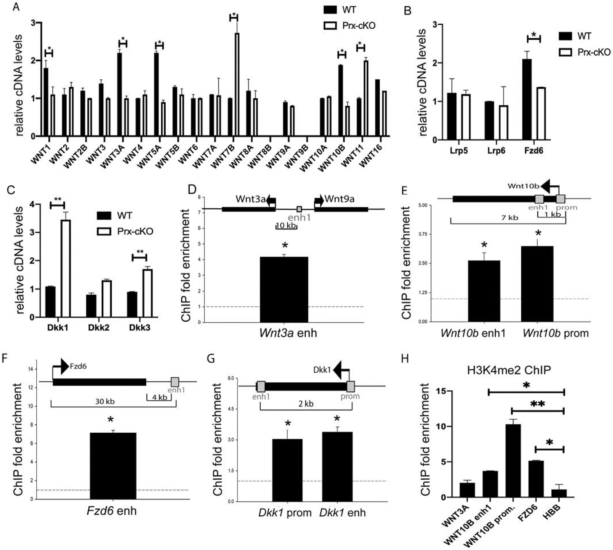
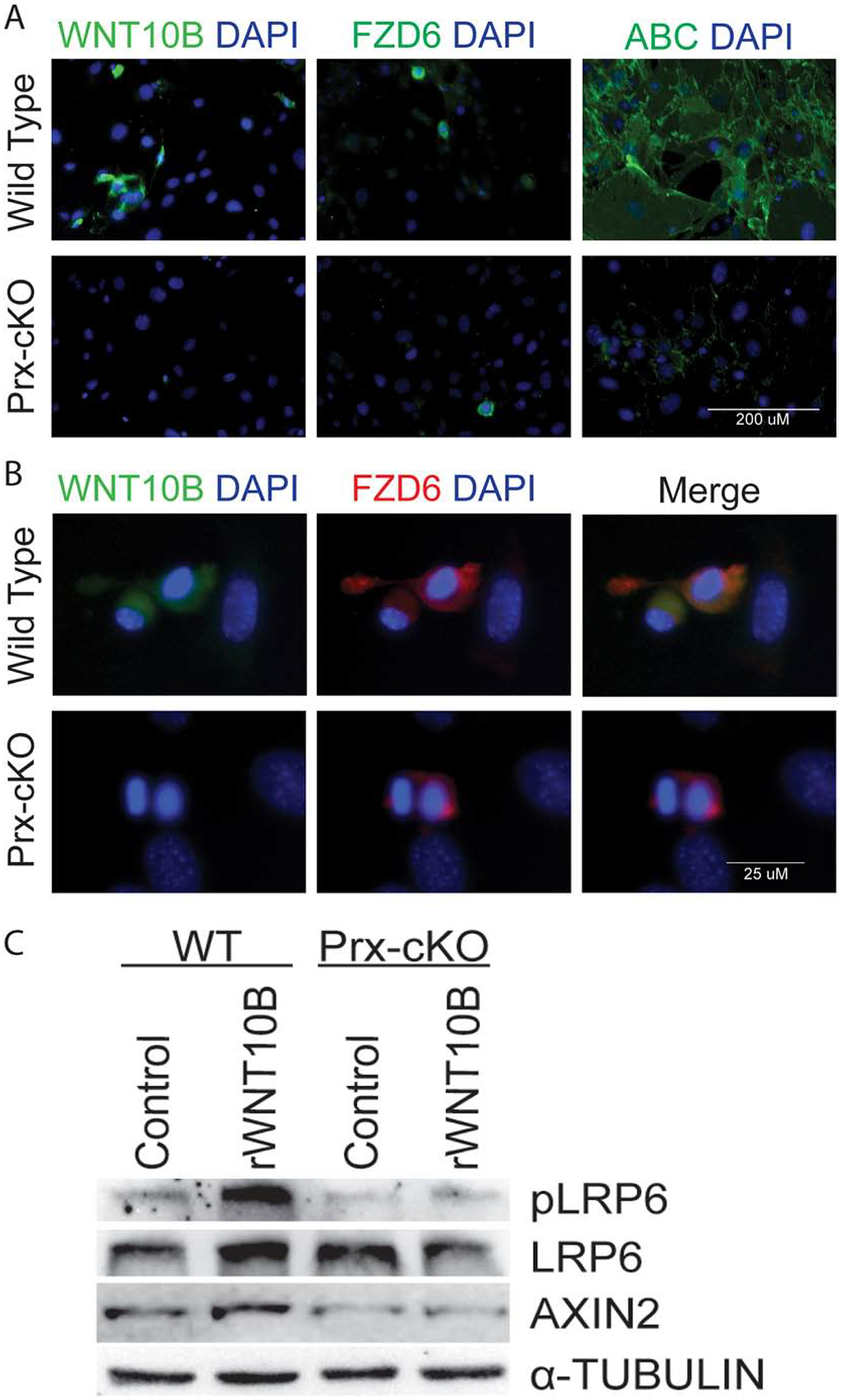
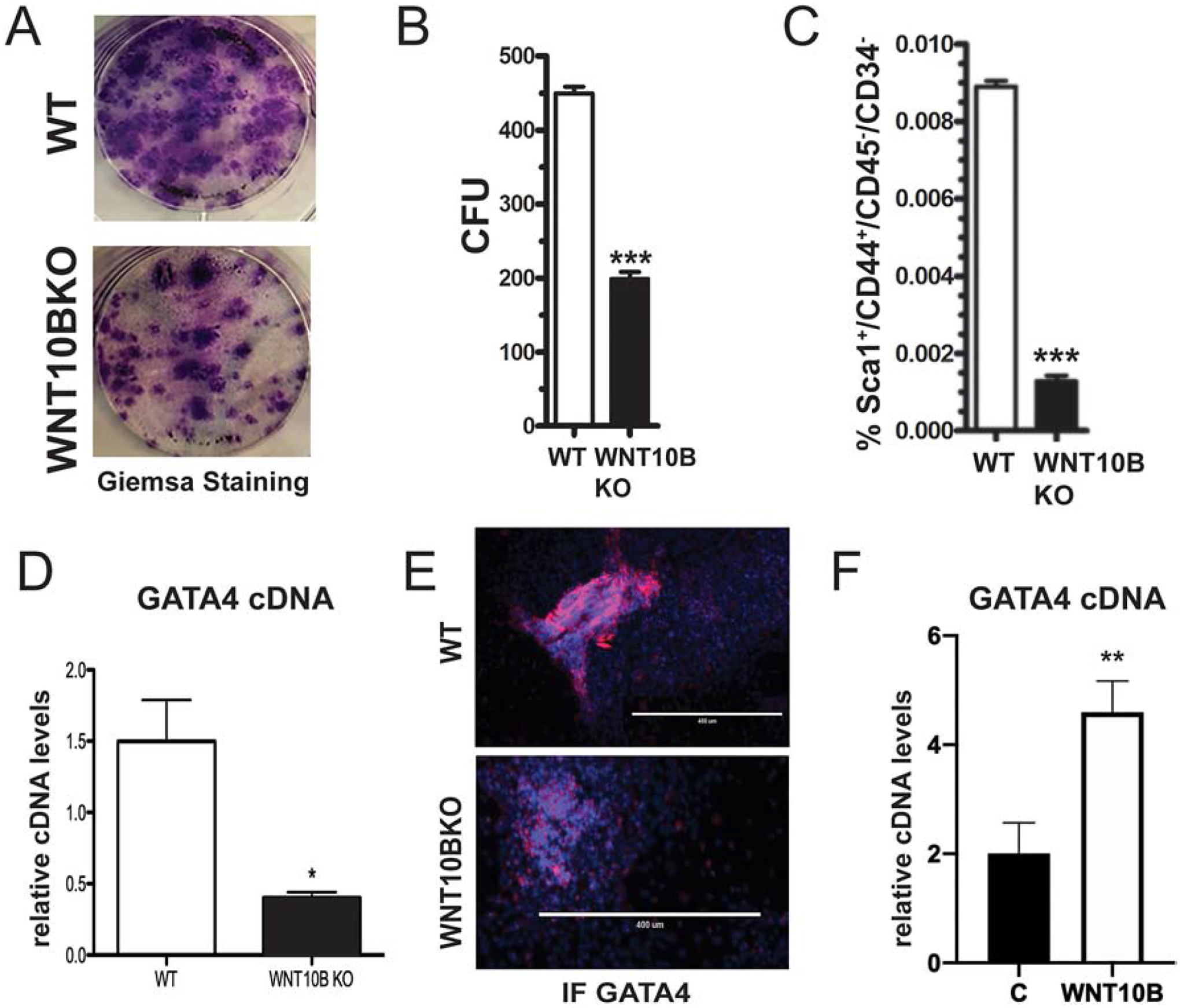
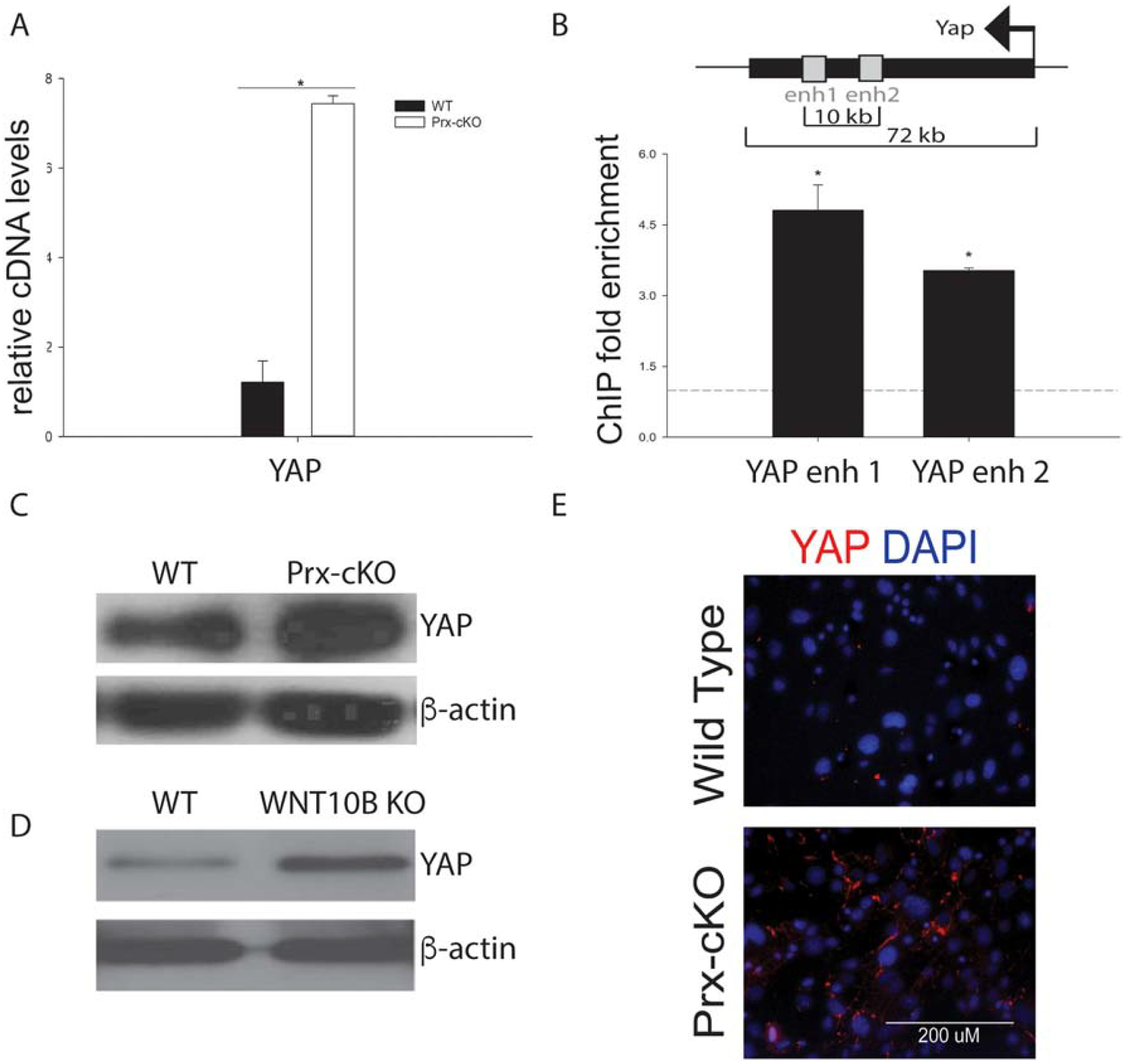
GATA4 is a transcription factor that regulates osteoblast differentiation. However, GATA4 is expressed at a higher level in mesenchymal stem cells (MSCs) than in osteoblasts. Therefore, the role of GATA4 in limb bud mesenchyme differentiation was investigated in mice by knocking out Gata4 using Cre-recombinase controlled by the Prx1 promoter (herein called Gata4 Prx-cKO mice). μCT analysis of the Gata4 Prx-cKO mice showed a decrease in trabecular bone properties compared with wildtype (Gata4fl/fl) littermates. Gata4 Prx-cKO mice have fewer MSCs as measured by CFU-F assays, mesenchymal progenitor cells (MPC2) (flow cytometry of Sca1+/CD45-/CD34-/CD44hi) and nestin immunofluorescence. Gata4 Prx-cKO bone marrow-derived MSCs have a significant reduction in WNT ligands, including WNT10B, and WNT signalosome components compared to control cells. Chromatin immunoprecipitation demonstrates that GATA4 is recruited to enhancers near Wnt3a, Wnt10b, Fzd6 and Dkk1. GATA4 also directly represses YAP in wildtype cells, and the absence of Gata4 leads to increased YAP expression. Together, we show that the decrease in MSCs is due to loss of Gata4 and a WNT10B-dependent positive autoregulatory loop. This leads to a concurrent increase of YAP and less activated β-catenin. These results explain the decreased trabecular bone in Gata4 Prx-cKO mice. We suggest that WNT signalosome activity in MSCs requires Gata4 and Wnt10b expression for lineage specification.

Keywords: GATA4; Mesenchymal stem cell; Osteoblast; WNT signaling; WNT10B.


Conflict of interest statement

Disclosure Statement: The authors declare that they have no conflict of interest.

Figures

Figure 1:
Figure 1:. Trabecular bone volume is decreased in Gata4 Prx-cKO mice.
(A, B) μCT reconstruction of trabecular bone in the femur of 14-week-old female mice. (C) Mean percentage bone volume (BV/TV), (D) trabecular number (Tb.N), (E) trabecular separation (Tb.Sp), (F) trabecular thickness (Tb.Th), (G) cortical thickness and (H) cortical porosity of WT (Gata4FL/FL) and Prx-cKO mice. Black bars indicate wildtype (WT) mice; white bars indicate GATA4 Prx-cKO mice. Data are mean ± standard deviation from 12 WT and 12 cKO mice. Student’s t-test: *P < 0.05 compared with WT.
Figure 2:
Figure 2:. GATA4 regulates both osteogenesis and adipogenesis in vitro.
Bone marrow MSCs from WT and Gata4 Prx-cKO were differentiated in osteogenic media for two weeks. (A) RNA was obtained and qPCR was performed for Gata4 and normalized to actin mRNA. (B) Cells were fixed and stained for mineralization using Alizarin Red. (C) Alizarin Red from part B was eluted and the mineral content measured at an OD of 570 nm. (D) qPCR was performed for Runx2 and (E) osteocalcin (OCN, BGLAP). (F) Bone marrow MSCs were also differentiated for two weeks in adipogenic media and qPCR was performed for Gata4 normalized to actin mRNA. (G) Adipocytes were stained using Oil Red O and (H) the percentage of fat droplets were calculated using ImageJ. (I) PPARγ and (J) Adiponectin expression was evaluated in the WT and Prx-cKO adipocytes. Student’s t test; * P < 0.05; *** P < 0.001 compared with wild-type. OD = optical density.
Figure 3:
Figure 3:. GATA4 regulates the number of mesenchymal stem cells.
(A-C) Bone marrow cells from WT and Gata4 Prx-cKO mice were cultured in MesenCult media. (A) Gata4 mRNA expression levels in undifferentiated cells from WT and Gata4 Prx-cKO mice. (B) Cells were stained with Giemsa and (C) the number of Giemsa colonies in multiple wells was quantified using ImageJ. (D and E) MSCs were analyzed for the expression of Sca1+/CD45−/CD34−/CD44 markers using flow cytometry. (F) The number of Sca1+/CD45−/CD34−/CD44lo (MPC1) cells and Sca1+/CD45−/CD34−/CD44hi (MPC2) from D and E are graphed. (G) Nestin expression was measured by qPCR from cells grown as in parts A-C. (H) Immunofluorescence of nestin in WT and Gata4 Prx-cKO mice (I) Percentage of nestin positive cells in three replicates as in part H. Student’s t test; * P < 0.05; *** P < 0.001 compared with wild-type.
Figure 4:
Figure 4:. GATA4 directly regulates WNT signaling.
Bone marrow cells from WT and Gata4 Prx-cKO mice were cultured in MesenCult media. RNA was collected and qPCR was performed to the indicated (A) Wnt genes, (B) Wnt Co-receptors, and (C) Wnt signaling inhibitors. (D-G) Chromatin immunoprecipitation was performed at the indicated enhancers or promoters. Streptavidin-coated beads were used to precipitate Flag-biotin-GATA4. qPCR was performed with primers to the indicated regions (gray boxes). Each PCR was normalized to input and represented as fold enrichment over a negative genomic locus (dotted line). Insets show a schematic of the genomic region. Black bars indicate genes with arrows indicating the start and direction of transcription. Gray boxes indicate enhancer or promoter regions. (H) ChIP with antibodies to H3K4me2 was performed and qPCR was performed with primers to the indicated regions. Student’s t test; * P < 0.05; ** P < 0.01, *** P < 0.001 compared with wild-type (A-C) or negative genomic locus (D-H).
Figure 5:
Figure 5:. WNT signaling is attenuated in GATA4 Prx-cKO mice.
(A) Bone marrow cells from WT and Gata4 Prx-cKO mice were cultured in MesenCult media and then fixed and stained with antibodies to WNT10B (green), FZD (green) or activated β-catenin (green). Nuclei were identified with DAPI (blue). (B) Bone marrow cells from WT and Gata4 Prx-cKO mice were cultured in MesenCult media and then fixed and stained with antibodies to WNT10B (green) or FZD6 (red). Nuclei were identified with DAPI (blue). (C) Bone marrow cells from WT and Gata4 Prx-cKO mice were cultured in MesenCult media. Cells were treated with 100 ng/mL rWNT10B for 30 minutes and then lysed for protein. Immunoblots were performed with antibodies to pLRP6, LRP6, AXIN2 and α-tubulin.
Figure 6:
Figure 6:. WNT10B regulates GATA4 expression.
(A) Bone marrow cells from WT and WNT10BKO mice were cultured in MesenCult media. Cells were stained with Giemsa and (B) the number of Giemsa colonies (CFU) was quantified using ImageJ. (C) MSCs were analyzed for the expression of Sca1+/CD45−/CD34−/CD44+ markers using flow cytometry. (D) Bone marrow cells from WT and WNT10BKO mice were cultured in MesenCult media. Gata4 mRNA expression levels were analyzed by qPCR in undifferentiated cells from WT and WNT10BKO mice. (E) Bone marrow cells from WT and WNT10BKO mice were cultured in MesenCult media. Cells were fixed and stained with an antibody to GATA4 (pink). Nuclei were identified with DAPI (blue). (F) WT osteoblasts were untreated or treated with 100 ng/mL rWNT10B for 24 hours and then lysed for RNA. GATA4 mRNA was detected by qPCR.
Figure 7:
Figure 7:. GATA4 directly represses YAP expression in MSCs.
(A) Bone marrow cells from WT and Gata4 Prx-cKO mice were cultured in MesenCult media. YAP cDNA expression levels were analyzed by qPCR in undifferentiated cells from WT and Gata4 Prx-cKO. (B) Chromatin immunoprecipitation was performed at the indicated enhancers or promoters. Streptavidin-coated beads were used to precipitate Flag-biotin-GATA4. qPCR was performed with primers to the indicated regions (gray boxes). Each PCR was normalized to input and represented as fold enrichment over a negative genomic locus (dotted line). Inset shows a schematic of the genomic region. The black bar indicates the YAP gene with the arrow indicating the start and direction of transcription. Gray boxes indicate intronic enhancer regions. Student’s t test; * P < 0.05; compared with a negative genomic locus. (C) Immunoblot for YAP and beta-actin from WT and Prx-cKO bone marrow. (D) Immunoblot for YAP and beta-actin from WT and WNT10B KO bone marrow. (E) Bone marrow cells from WT and Gata4 Prx-cKO mice were cultured in MesenCult media. Cells were fixed and stained with an antibody to YAP (red). Nuclei were identified with DAPI (blue).

References

    1. Guemes M, Garcia AJ, Rigueur D, Runke S, Wang W, Zhao G, Mayorga VH, Atti E, Tetradis S, Peault B, Lyons K, Miranda-Carboni GA, and Krum SA, GATA4 is essential for bone mineralization via ERalpha and TGFbeta/BMP pathways. J Bone Miner Res, 2014. 29(12): p. 2676–87. - PMC - PubMed
    1. Miranda-Carboni GA, Guemes M, Bailey S, Anaya E, Corselli M, Peault B, and Krum SA, GATA4 regulates estrogen receptor-alpha-mediated osteoblast transcription. Mol Endocrinol, 2011. 25(7): p. 1126–36. - PMC - PubMed
    1. Khalid AB, Slayden AV, Kumpati J, Perry CD, Lillo MA, Arroyo SR, Miranda-Carboni G, and Krum SA, GATA4 directly regulates Runx2 expression and osteoblast differentiation JBMR Plus, 2018. 2(2): p. 81–91. - PMC - PubMed
    1. Khalid AB, Slayden AV, Kumpati J, Perry CD, Berryhill SB, Crawford JA, Fatima I, Morselli M, Pellegrini M, Miranda-Carboni GA, and Krum SA, GATA4 represses RANKL in osteoblasts via multiple long-range enhancers to regulate osteoclast differentiation. Bone, 2018. 116: p. 78–86. - PMC - PubMed
    1. Sera SR and Zur Nieden NI, microRNA Regulation of Skeletal Development. Curr Osteoporos Rep, 2017. 15(4): p. 353–366. - PMC - PubMed

Publication types