Review
. 2020 Dec 16;18(12):645.
doi: 10.3390/md18120645.

Bioactivity Potential of Marine Natural Products from Scleractinia-Associated Microbes and In Silico Anti-SARS-COV-2 Evaluation

Affiliations
Review

Bioactivity Potential of Marine Natural Products from Scleractinia-Associated Microbes and In Silico Anti-SARS-COV-2 Evaluation

Eman Maher Zahran et al. Mar Drugs. .

Abstract

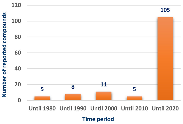
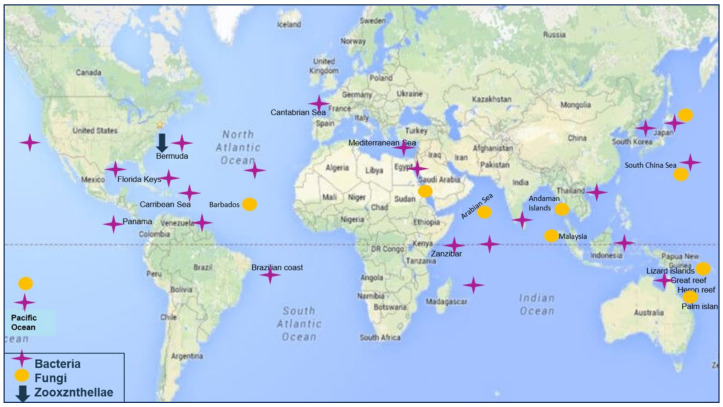
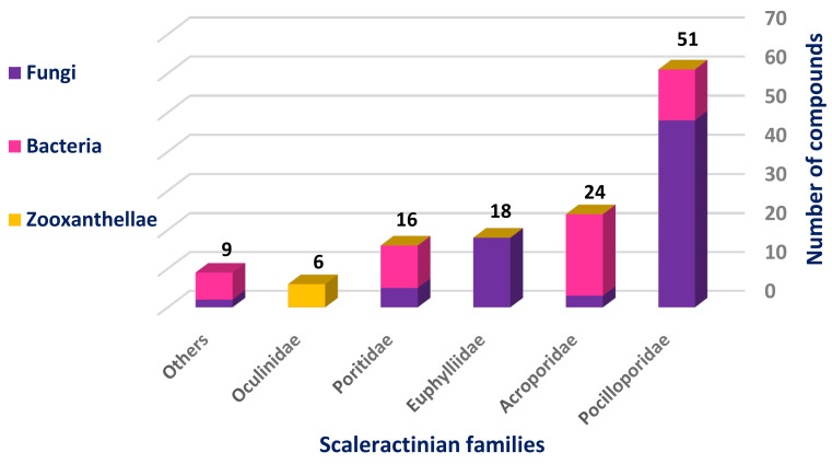
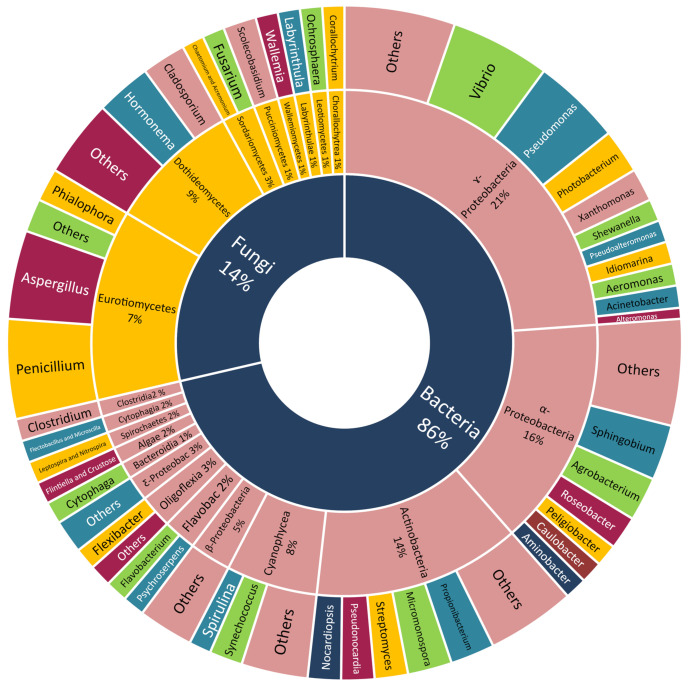
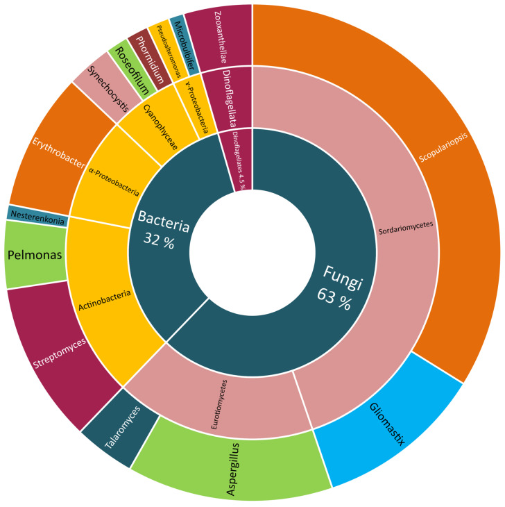
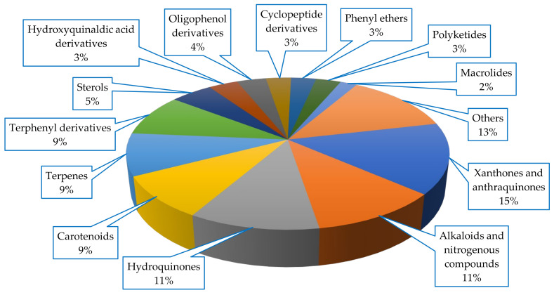
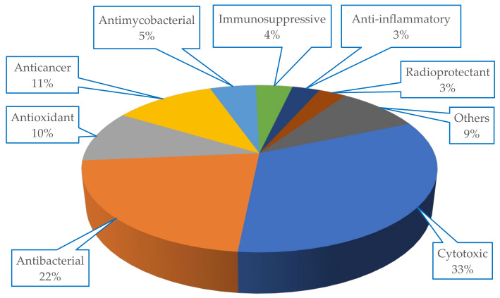
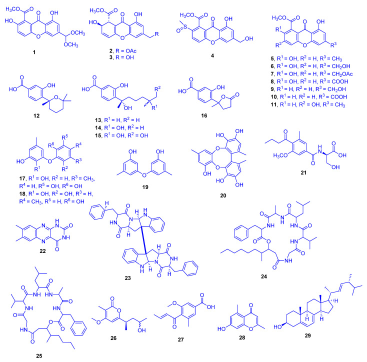
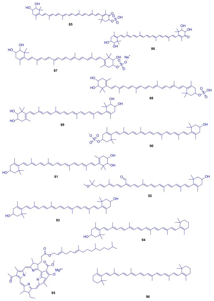
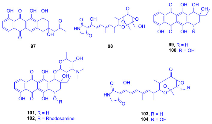
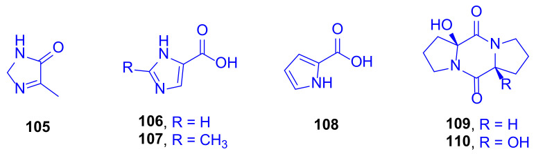
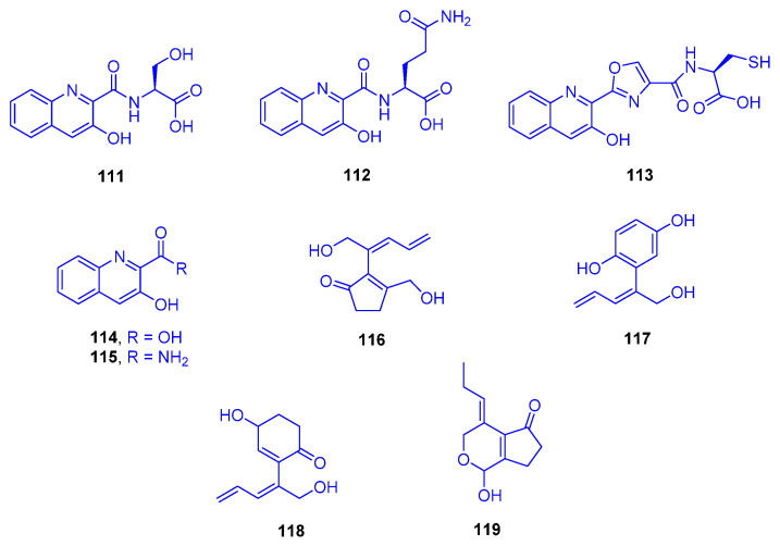
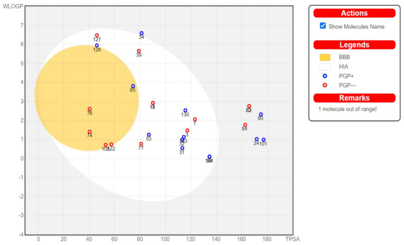
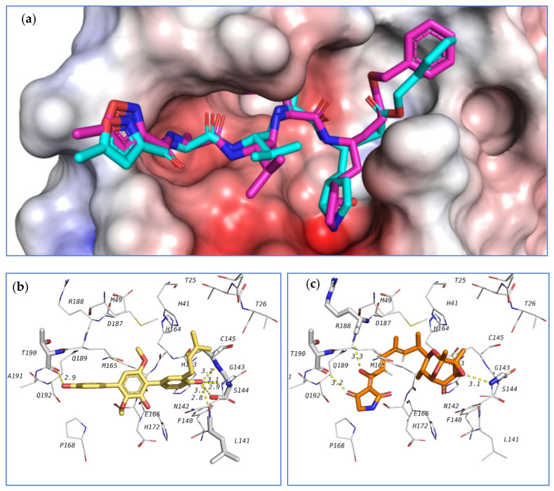
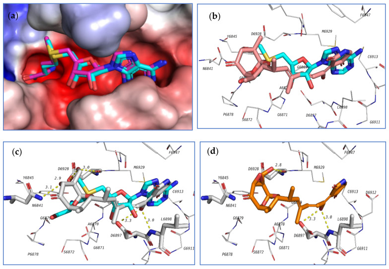
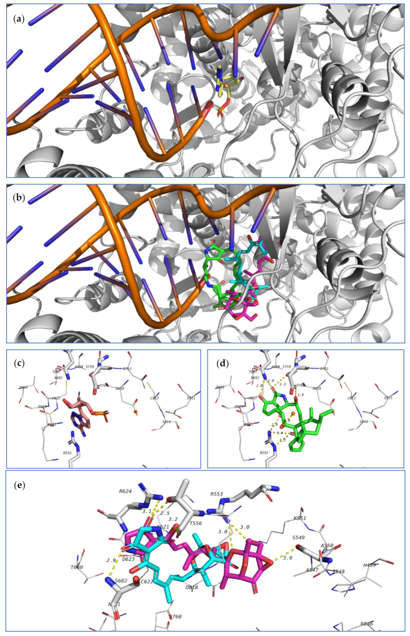
Marine organisms and their associated microbes are rich in diverse chemical leads. With the development of marine biotechnology, a considerable number of research activities are focused on marine bacteria and fungi-derived bioactive compounds. Marine bacteria and fungi are ranked on the top of the hierarchy of all organisms, as they are responsible for producing a wide range of bioactive secondary metabolites with possible pharmaceutical applications. Thus, they have the potential to provide future drugs against challenging diseases, such as cancer, a range of viral diseases, malaria, and inflammation. This review aims at describing the literature on secondary metabolites that have been obtained from Scleractinian-associated organisms including bacteria, fungi, and zooxanthellae, with full coverage of the period from 1982 to 2020, as well as illustrating their biological activities and structure activity relationship (SAR). Moreover, all these compounds were filtered based on ADME analysis to determine their physicochemical properties, and 15 compounds were selected. The selected compounds were virtually investigated for potential inhibition for SARS-CoV-2 targets using molecular docking studies. Promising potential results against SARS-CoV-2 RNA dependent RNA polymerase (RdRp) and methyltransferase (nsp16) are presented.

Keywords: ADME analysis; RNA-dependent RNA polymerase; SARS-CoV-2; Scleractinia; marine bacteria; marine fungi; marine natural products; methyltransferase; molecular docking; zooxanthellae.


Conflict of interest statement

The authors declare no conflict of interest.

Figures

Figure 1
Figure 1
Number of compounds isolated from Scleractinia-associated organisms according to the year of publication.
Figure 2
Figure 2
Geographical distribution of isolated hard corals-associated organisms.
Figure 3
Figure 3
Number of compounds isolated from different studied Scleractinian families-associated organisms.
Figure 4
Figure 4
Scleractinia-associated organisms studied genera without isolated compounds.
Figure 5
Figure 5
Scleractinia-associated organisms studied genera with isolated compounds.
Figure 6
Figure 6
Chemical skeletons of compounds isolated from Scleractinia-associated organisms.
Figure 7
Figure 7
Biological activity spectrum of compounds isolated from Scleractinia-associated organisms.
Figure 8
Figure 8
12-Dimethoxypinselin (1), 12-O-acetyl-AGI-B4 (2), AGI-B4 (3), hyperxanthone C (4), pinselin (5), sydowinin B (6), 13-O-acetyl-sydowinin B (7), compound 8, sydowinin A (9), compound 10, compound 11, sydowic acid (12), sydonic acid (13), 11-hydroxysydonic acid (14), 11,12-dihydroxysydonic acid (15), 1-hydroxyboivinianic acid (16), violaceol I (17), violaceol II (18), diorcinol (19), rikuzenol (20), scopulamide (21), lumichrome (22), WIN 64821 (23), scopularide A (24), scopularide B (25), scopupyrone (26), pyrenochaetic acid (27), 7-OH-2,5-dimethylchromone (28) and ergosterol (29).
Figure 9
Figure 9
4-Methylcandidusin A (30), aspetritone A (31), aspetritone B (32), 3,4-dimethyl-3″-prenylcandidusin A (33), 4-methyl-3″-prenylcandidusin A (34), 3,4-dimethylcandidusin A (35), candidusin A (36), 4,4′-deoxyterphenyllin (37), 4″-deoxyterphenyllin (38), 3-prenylterphenyllin (39), terphenyllin (40), 3-hydroxyterphenyllin (41), 3-hydroxy-3″-deoxyterphenyllin (42), 3″-prenyl-terphenyllin (43), emodin (44) and compounds 4547.
Figure 10
Figure 10
3β,7β,15α,24-Tetrahydroxyolean-12-ene-11,22-dione (48), 15α,22β,24-trihydroxyolean-11,13-diene-3-one (49), 7β,15α,24-trihydroxyolean-12-ene-3,11,22-trione (50), 15α,24-dihydroxyolean-12-ene-3,11,22-trione (51), soyasapogenol B (52), (2E, 4E)-4′-dihydrophaseic acid (53), (2Z, 4E)-4′-dihydrophaseic acid (54), 6-hydroxy-2,7-dimethyl-1,4-naphthoquinone (55), 6-hydroxy-2,2-dimethyl-2H-chromene (56), scoparone (57), 5-methyluracil (58) and compounds 5964.
Figure 11
Figure 11
Gliomastin A (65), gliomastin B (66), gliomastin C (67), gliomastin D (68), 9-O-methylgliomastin C (69), acremonin A 1-O-β-D-glucopyranoside (70), gliomastin E 1-O- β-D-glucopyranoside (71), 6′-O-acetyl-isohomoarbutin (72), isohomoarbutin (73), 2-methyl-1,4-benzenediol (74), acremonin A (75), prenylhydroquinone (76), F-11334A1 (77), compound 78 and compound 79.
Figure 12
Figure 12
Verruculosin A (80), verruculosin B (81), bacillisporin F (82), duclauxin (83) and xenoclauxin (84).
Figure 13
Figure 13
Erythroxanthin sulfate (85), ketonostoxanthin (86), nostoxanthin sulfate (87), caloxanthin sulfate (88), nostoxanthin (89), zeaxanthin sulfate (90), caloxanthin (91), bacteriorubixanthinal (92), zeaxanthin (93), β-cryptoxanthin (94), bacteriochlorophyll (95) and β-carotene (96).
Figure 14
Figure 14
Aranciamycin K (97), isotirandamycin B (98), γ-rhodomycinone (99), β-rhodomycinone (100), 262-6 (101), β-rhodomycin-II (102), tirandamycin A (103) and tirandamycin B (104).
Figure 15
Figure 15
Pelopuradazole (105), 3H-imidazole-4-carboxylic acid (106), 2-methyl-3H-imidazole-4-carboxylic acid (107), 1H-pyrrole-2-carboxylic acid (108), pelopurin A (109) and pelopurin B (110).
Figure 16
Figure 16
Compounds 111113, 3-hydroxyquinaldic acid (114), 3-hydroxyquinaldic acid amide (115), nakienone A (116), nakitriol (117), nakienone B (118) and nakienone C (119).
Figure 17
Figure 17
Pitiamide A (120), 1E-pitiamide B (121), looekeyolide A (122), looekeyolide B (123), alteramide A (124), alteramide B (125), (2Z,4E)-3-methyl-2,4-decadienoic acid (126), nesteretal A (127) and lobophorin K (128).
Figure 18
Figure 18
Gorgosterol (129), 23-desmethyl-gorgosterol (130), dinosterol (131), cholesterol (132), 4α-(24S)-dimethyl-cholesta-3β-ol (133) and 4α-(24R)-dimethyl-cholesta-22-en-3β-ol (134).
Figure 19
Figure 19
Boiled-egg diagram of the investigated compounds. BBB, passively permeate blood brain barrier; HIA, Human intestinal absorption; PGP+, compounds that effluated from central nervous system by the P-Glycoprotein; PGP-, compounds that is not binding to P-Glycoprotein.
Figure 20
Figure 20
Docking results of investigated compounds in the active site of SARS-CoV-2 main protease (6LU7). (a) Validation of docking procedure showing good matching between crystallized (blue) and docked (pink) ligands. (b) Docking pose and interactions of 39 (yellow). (c) Docking pose and interactions of 103 (orange).
Figure 21
Figure 21
Docking results of the investigated compounds in the active site of SARS-CoV-2 methyltransferase (6W4H). (a) Validation of the docking procedure showing good matching between crystallized (blue) and docked (pink) ligands. (b) Docking pose and interactions of 103 (brick-red). (c) Docking pose and interactions of 98 (white). (d) Docking pose and interactions of 104 (orange).
Figure 22
Figure 22
Docking results of the tested compounds in the active site of SARS-CoV-2 RdRp (7BV2). (a) Binding position of remdesivir. (b) Binding position of the docked ligands including 124 (green), 103 (blue), and 104 (pink). (c) Interactions between co-crystallized ligand with amino acids in the active site of RdRp. (d) Interactions of 124 (green). (e) Interactions of 103 (blue) and 104 (pink).

References

    1. El-Hossary E.M., Cheng C., Hamed M.M., Hamed A.N.E.-S., Ohlsen K., Hentschel U., Abdelmohsen U.R. Antifungal potential of marine natural products. Eur. J. Med. Chem. 2017;126:631–651. doi: 10.1016/j.ejmech.2016.11.022. - DOI - PubMed
    1. Liu M., El-Hossary E.M., Oelschlaeger T.A., Donia M.S., Quinn R.J., Abdelmohsen U.R. Potential of marine natural products against drug-resistant bacterial infections. Lancet Infect. Dis. 2019;19:e237–e245. doi: 10.1016/S1473-3099(18)30711-4. - DOI - PubMed
    1. Shady N.H., El-Hossary E.M., Fouad M.A., Gulder T.A.M., Kamel M.S., Abdelmohsen U.R. Bioactive Natural Products of Marine Sponges from the Genus Hyrtios. Molecules. 2017;22:781. doi: 10.3390/molecules22050781. - DOI - PMC - PubMed
    1. Abdelmohsen U.R., Balasubramanian S.A., Oelschlaeger T., Grkovic T., Pham N.B., Quinn R.J., Hentschel U. Potential of marine natural products against drug-resistant fungal, viral, and parasitic infections. Lancet Infect. Dis. 2017;17:e30–e41. doi: 10.1016/S1473-3099(16)30323-1. - DOI - PubMed
    1. Abdelmohsen U.R., Bayer K., Hentschel U. Diversity, abundance and natural products of marine sponge-associated actinomycetes. Nat. Prod. Rep. 2014;31:381–399. doi: 10.1039/C3NP70111E. - DOI - PubMed