Review
. 2020 Dec 16;10(12):1682.
doi: 10.3390/biom10121682.

S-adenosyl-l-homocysteine Hydrolase: A Structural Perspective on the Enzyme with Two Rossmann-Fold Domains

Affiliations
Review

S-adenosyl-l-homocysteine Hydrolase: A Structural Perspective on the Enzyme with Two Rossmann-Fold Domains

Krzysztof Brzezinski. Biomolecules. .

Abstract

S-adenosyl-l-homocysteine hydrolase (SAHase) is a major regulator of cellular methylation reactions that occur in eukaryotic and prokaryotic organisms. SAHase activity is also a significant source of l-homocysteine and adenosine, two compounds involved in numerous vital, as well as pathological processes. Therefore, apart from cellular methylation, the enzyme may also influence other processes important for the physiology of particular organisms. Herein, presented is the structural characterization and comparison of SAHases of eukaryotic and prokaryotic origin, with an emphasis on the two principal domains of SAHase subunit based on the Rossmann motif. The first domain is involved in the binding of a substrate, e.g., S-adenosyl-l-homocysteine or adenosine and the second domain binds the NAD+ cofactor. Despite their structural similarity, the molecular interactions between an adenosine-based ligand molecule and macromolecular environment are different in each domain. As a consequence, significant differences in the conformation of d-ribofuranose rings of nucleoside and nucleotide ligands, especially those attached to adenosine moiety, are observed. On the other hand, the chemical nature of adenine ring recognition, as well as an orientation of the adenine ring around the N-glycosidic bond are of high similarity for the ligands bound in the substrate- and cofactor-binding domains.

Keywords: cellular methylation; nucleoside substrate; nucleotide cofactor; protein structure; protein-ligand interactions; structural enzymology.


Conflict of interest statement

The author declares no conflict of interest.

Figures

Figure 1
Figure 1
Oligomeric forms of SAHases; (a) The AB (or A’B’) homodimer corresponds to the active form of the plant enzyme; (b) The dimer of dimers (AB–A’B’) that corresponds to the active form of most SAHases of archaeal, bacterial and eukaryotic origin. The colour code indicates individual subunits: A and A’ (blue), B and B’ (red).
Figure 2
Figure 2
A topology diagram of the Rossmann fold in (a) the substrate-binding domain and (b) the cofactor-binding domain. Secondary structure elements present in the canonical Rossmann motif are shown in yellow and red. Additional, non-canonical β-chains present in all SAHases, in the central β-sheet, are shown in grey. The secondary structure elements present in the substrate binding-domain (SBD) of plant and numerous bacterial SAHases are shown in green. Other, variable secondary structure elements are omitted (broken lines). The numbering scheme of the secondary structure elements is based on their order in the polypeptide chain of the Rossmann fold-based domains of SAHases. For those elements that are absent in the canonical Rossmann fold, the letter designation is used. Amino acid residue numbers correspond to those of LlSAHase.
Figure 3
Figure 3
Multiple sequence alignment of selected SAHases. Residues on the yellow background are involved in a ligand binding in the substrate-binding domain, while residues on the blue background interact with the NAD+ cofactor molecule in the cofactor-binding domain. Two specific motifs involved in a ligand binding are highlighted. Amino acid residue numbers correspond to those of LlSAHase. The alignment includes the sequences of the following species (UniProt accession code): Lupinus luteus (Q9SP37), Plasmodium falciparum (P50250), Burkholderia pseudomallei (Q3JY79), Mycobacterium tuberculosis (P9WGV3), Pseudomonas aeruginosa (Q9I685), Cytophaga hutchinsonii (A0A6N4SNR7), Trypanosoma brucei (Q383X0), Homo sapiens (P23526), Saccharolobus solfataricus (P50252), Archaeoglobus fulgidus (O28279), and Thermotoga maritima (O51933).
Figure 4
Figure 4
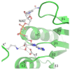
The canonical mode of NAD+ binding in the Rossmann fold-based CBD of SAHases. Residues located in the region of loop β1 and the N-terminal end of α1 helix, as well as the acidic residue from the β2 chain, are involved in the interactions with the cofactor molecule. Dashed lines indicate potential hydrogen bonds between 2′- and 3′-hydroxyl groups of the adenosine moiety and the conserved glutamate residue (E) from subunit A, and the conserved lysine residue from DD from the adjacent subunit B (KB).
Figure 5
Figure 5
The binding mode of ligands present in the SBD domain of plant SAHase from Lupinus luteus: (a) adenosine (Ado), (b) 3′-deoxyadenosine (3′-dAdo), and (c) 2′-deoxyadenosine [26]. The potential polar interactions are marked with red dashed lines. Residues from the CBD domain are marked with stars. Water molecules are shown as red spheres.
Figure 6
Figure 6
The recognition mode of the purine ring in (a) the substrate-binding domain and (b) the cofactor-binding domain of plant SAHase from Lupinus luteus.
Figure 7
Figure 7
The recognition mode of the nicotinamide riboside moiety of the bound cofactor in plant SAHase from Lupinus luteus.
Figure 8
Figure 8
Accessibility of the active site from the solvent region is regulated by a “molecular gate” formed by a conserved His-Phe diad; (a) For the rat enzyme in its substrate-free state the channel is open, whereas (b) for the human enzyme in a complex with the adenosine analog (2′-hydroxy- 3′-ketocyclopent-4′-enyladenine, DHCeA), the channel is closed with no access to the substrate-binding site. Amino acid residue numbers correspond to those of human and rat SAHase.

References

    1. Rossmann M.G., Moras D., Olsen K.W. Chemical and biological evolution of nucleotide-binding protein. Nature. 1974;250:194–199. doi: 10.1038/250194a0. - DOI - PubMed
    1. Aravind L., Mazumder R., Vasudevan S., Koonin E.V. Trends in protein evolution inferred from sequence and structure analysis. Curr. Opin. Struct. Biol. 2002;12:392–399. doi: 10.1016/S0959-440X(02)00334-2. - DOI - PubMed
    1. Xie L., Bourne P.E. Detecting evolutionary relationships across existing fold space, using sequence order-independent profile-profile alignments. Proc. Natl. Acad. Sci. USA. 2008;105:5441–5446. doi: 10.1073/pnas.0704422105. - DOI - PMC - PubMed
    1. Edwards H., Abeln S., Deane C.M. Exploring fold space preferences of new-born and ancient protein superfamilies. PLoS Comput. Biol. 2013;9 doi: 10.1371/journal.pcbi.1003325. - DOI - PMC - PubMed
    1. Bukhari S.A., Caetano-Anollés G. Origin and evolution of protein fold designs inferred from phylogenomic analysis of CATH domain structures in proteomes. PLoS Comput. Biol. 2013;9:e1003009. doi: 10.1371/journal.pcbi.1003009. - DOI - PMC - PubMed

Publication types