When pneumonia is not COVID-19
- PMID: 33339621
- PMCID: PMC7813497
- DOI: 10.1016/j.rx.2020.11.003
When pneumonia is not COVID-19
Abstract
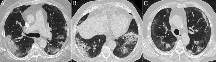
During the COVID-19 epidemic, the prevalence of the disease means that practically any lung opacity on an X-ray could represent pneumonia due to infection with SARS-CoV-2. Nevertheless, atypical radiologic findings add weight to negative microbiological or serological tests. Likewise, outside the epidemic wave and with the return of other respiratory diseases, radiologists can play an important role in decision making about diagnoses, treatment, or preventive measures (isolation), provided they know the key findings for entities that can simulate COVID-19 pneumonia. Unifocal opacities or opacities located in upper lung fields and predominant airway involvement, in addition to other key radiologic and clinical findings detailed in this paper, make it necessary to widen the spectrum of possible diagnoses.
La prevalencia en fase epidémica de la COVID-19 hace que prácticamente cualquier opacidad pulmonar en la radiografía de tórax pueda ser una neumonía por SARS-CoV-2. Sin embargo, hallazgos radiológicos atípicos aumentarán la credibilidad de un resultado microbiológico o serológico negativo. Asimismo, fuera de la ola epidémica y con el retorno de otras entidades respiratorias, el radiólogo puede tener gran relevancia en la toma de decisiones diagnósticas, terapéuticas o preventivas (aislamiento) si conoce las claves diagnósticas de las entidades simuladoras de neumonía COVID-19. La distribución unifocal o en campos pulmonares superiores de las opacidades y la afectación predominante de vía aérea, entre otras claves radiológicas y clínicas detalladas en este capítulo, implican necesariamente ampliar el abanico de posibilidades diagnósticas.
Keywords: COVID-19; Computed tomography; Diagnostic imaging; Radiography; X ray; X-rays.
Copyright © 2020 SERAM. Publicado por Elsevier España, S.L.U. All rights reserved.
Figures
Similar articles
-
Radiologic diagnosis of patients with COVID-19.Radiologia (Engl Ed). 2021 Jan-Feb;63(1):56-73. doi: 10.1016/j.rx.2020.11.001. Epub 2020 Nov 24. Radiologia (Engl Ed). 2021. PMID: 33339622 Free PMC article.
-
Chest-CT mimics of COVID-19 pneumonia-a review article.Emerg Radiol. 2021 Jun;28(3):507-518. doi: 10.1007/s10140-021-01919-0. Epub 2021 Mar 1. Emerg Radiol. 2021. PMID: 33646498 Free PMC article. Review.
-
COVID-19 pneumonia-ultrasound, radiographic, and computed tomography findings: a comprehensive pictorial essay.Emerg Radiol. 2021 Jun;28(3):519-526. doi: 10.1007/s10140-021-01905-6. Epub 2021 Jan 30. Emerg Radiol. 2021. PMID: 33517546 Free PMC article. Review.
-
Chest radiography findings of COVID-19 pneumonia: a specific pattern for a confident differential diagnosis.Acta Radiol. 2022 Dec;63(12):1619-1626. doi: 10.1177/02841851211055163. Epub 2021 Nov 13. Acta Radiol. 2022. PMID: 34779269 Free PMC article.
-
COVID-19 Associated Pneumonia: A review of chest radiograph and computed tomography findings.Sultan Qaboos Univ Med J. 2021 Feb;21(1):e4-e11. doi: 10.18295/squmj.2021.21.01.002. Epub 2021 Mar 15. Sultan Qaboos Univ Med J. 2021. PMID: 33777418 Free PMC article. Review.
Cited by
-
Efficacy of Transfer Learning-based ResNet models in Chest X-ray image classification for detecting COVID-19 Pneumonia.Chemometr Intell Lab Syst. 2022 May 15;224:104534. doi: 10.1016/j.chemolab.2022.104534. Epub 2022 Mar 11. Chemometr Intell Lab Syst. 2022. PMID: 35291673 Free PMC article.
-
Comparison of pneumonia features in children caused by SARS-CoV-2 and other viral respiratory pathogens.Pediatr Pulmonol. 2022 Oct;57(10):2374-2382. doi: 10.1002/ppul.26042. Epub 2022 Jul 8. Pediatr Pulmonol. 2022. PMID: 35754093 Free PMC article.
-
Epidemiology of Systemic Mycoses in the COVID-19 Pandemic.J Fungi (Basel). 2021 Jul 13;7(7):556. doi: 10.3390/jof7070556. J Fungi (Basel). 2021. PMID: 34356935 Free PMC article. Review.
-
Delayed Diagnosis of an Atypical Pneumonia Resembling a Solitary Pulmonary Nodule.Cureus. 2021 Nov 10;13(11):e19456. doi: 10.7759/cureus.19456. eCollection 2021 Nov. Cureus. 2021. PMID: 34926029 Free PMC article.
-
Pneumonia in the Pandemic: Not Always COVID-19.Cureus. 2023 Oct 27;15(10):e47802. doi: 10.7759/cureus.47802. eCollection 2023 Oct. Cureus. 2023. PMID: 38021945 Free PMC article.
References
Publication types
MeSH terms
LinkOut - more resources
Full Text Sources
Other Literature Sources
Medical
Miscellaneous