

Abnormalities of synaptic mitochondria in autism spectrum disorder and related neurodevelopmental disorders
- PMID: 33340060
- PMCID: PMC7819932
- DOI: 10.1007/s00109-020-02018-2
Abnormalities of synaptic mitochondria in autism spectrum disorder and related neurodevelopmental disorders
Abstract
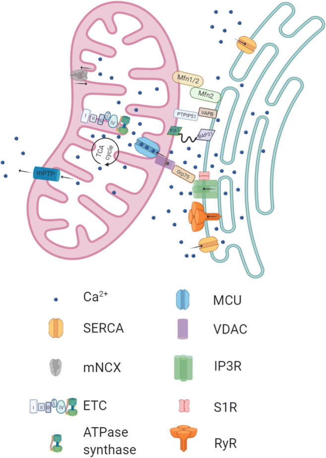
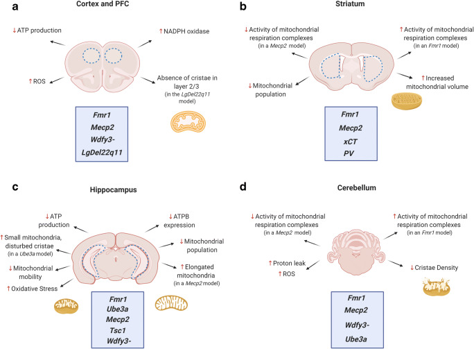
Autism spectrum disorder (ASD) is a neurodevelopmental condition primarily characterized by an impairment of social interaction combined with the occurrence of repetitive behaviors. ASD starts in childhood and prevails across the lifespan. The variability of its clinical presentation renders early diagnosis difficult. Mutations in synaptic genes and alterations of mitochondrial functions are considered important underlying pathogenic factors, but it is obvious that we are far from a comprehensive understanding of ASD pathophysiology. At the synapse, mitochondria perform diverse functions, which are clearly not limited to their classical role as energy providers. Here, we review the current knowledge about mitochondria at the synapse and summarize the mitochondrial disturbances found in mouse models of ASD and other ASD-related neurodevelopmental disorders, like DiGeorge syndrome, Rett syndrome, Tuberous sclerosis complex, and Down syndrome.
Keywords: ASD; Autism spectrum disorder; Mitochondria; Neurodevelopmental disorders; Synapse.
Conflict of interest statement
The authors declare that they have no conflict of interest.
Figures

Similar articles
-
Glutamatergic synapses in neurodevelopmental disorders.Prog Neuropsychopharmacol Biol Psychiatry. 2018 Jun 8;84(Pt B):328-342. doi: 10.1016/j.pnpbp.2017.09.014. Epub 2017 Sep 19. Prog Neuropsychopharmacol Biol Psychiatry. 2018. PMID: 28935587 Review.
-
NEXMIF/KIDLIA Knock-out Mouse Demonstrates Autism-Like Behaviors, Memory Deficits, and Impairments in Synapse Formation and Function.J Neurosci. 2020 Jan 2;40(1):237-254. doi: 10.1523/JNEUROSCI.0222-19.2019. Epub 2019 Nov 8. J Neurosci. 2020. PMID: 31704787 Free PMC article.
-
Plasticity of dendritic spines: Molecular function and dysfunction in neurodevelopmental disorders.Psychiatry Clin Neurosci. 2019 Sep;73(9):541-550. doi: 10.1111/pcn.12899. Epub 2019 Jul 8. Psychiatry Clin Neurosci. 2019. PMID: 31215705 Review.
-
Synaptopathology Involved in Autism Spectrum Disorder.Front Cell Neurosci. 2018 Dec 21;12:470. doi: 10.3389/fncel.2018.00470. eCollection 2018. Front Cell Neurosci. 2018. PMID: 30627085 Free PMC article. Review.
-
The Potential Role of AMPA Receptor Trafficking in Autism and Other Neurodevelopmental Conditions.Neuroscience. 2021 Dec 15;479:180-191. doi: 10.1016/j.neuroscience.2021.09.013. Epub 2021 Sep 24. Neuroscience. 2021. PMID: 34571086 Review.
Cited by
-
Association of NGF and Mitochondrial Respiration with Autism Spectrum Disorder.Int J Mol Sci. 2022 Oct 7;23(19):11917. doi: 10.3390/ijms231911917. Int J Mol Sci. 2022. PMID: 36233217 Free PMC article.
-
NR2F1 shapes mitochondria in the mouse brain, providing new insights into Bosch-Boonstra-Schaaf optic atrophy syndrome.Dis Model Mech. 2023 Jun 1;16(6):dmm049854. doi: 10.1242/dmm.049854. Epub 2023 Jun 26. Dis Model Mech. 2023. PMID: 37260288 Free PMC article.
-
Early Chronic Memantine Treatment-Induced Transcriptomic Changes in Wild-Type and Shank2-Mutant Mice.Front Mol Neurosci. 2021 Sep 14;14:712576. doi: 10.3389/fnmol.2021.712576. eCollection 2021. Front Mol Neurosci. 2021. PMID: 34594187 Free PMC article.
-
Camk2a-Cre and Tshz3 Expression in Mouse Striatal Cholinergic Interneurons: Implications for Autism Spectrum Disorder.Front Genet. 2021 Jul 12;12:683959. doi: 10.3389/fgene.2021.683959. eCollection 2021. Front Genet. 2021. PMID: 34349780 Free PMC article.
-
Increased expression of SLC25A1/CIC causes an autistic-like phenotype with altered neuron morphology.Brain. 2022 Apr 18;145(2):500-516. doi: 10.1093/brain/awab295. Brain. 2022. PMID: 35203088 Free PMC article.
References
-
- Muhle RA, Reed HE, Stratigos KA, Veenstra-VanderWeele J. The emerging clinical neuroscience of autism spectrum disorder: a review. JAMA Psychiatry. 2018;75(5):514–523. - PubMed
-
- Bourgeron T. From the genetic architecture to synaptic plasticity in autism spectrum disorder. Nat Rev Neurosci. 2015;16(9):551–563. - PubMed
Publication types
MeSH terms
LinkOut - more resources
Full Text Sources
