Review
. 2021 Mar;78(6):2621-2639.
doi: 10.1007/s00018-020-03722-5. Epub 2020 Dec 19.

Autophagy and the endolysosomal system in presynaptic function

Affiliations
Review

Autophagy and the endolysosomal system in presynaptic function

Maria Andres-Alonso et al. Cell Mol Life Sci. 2021 Mar.

Abstract

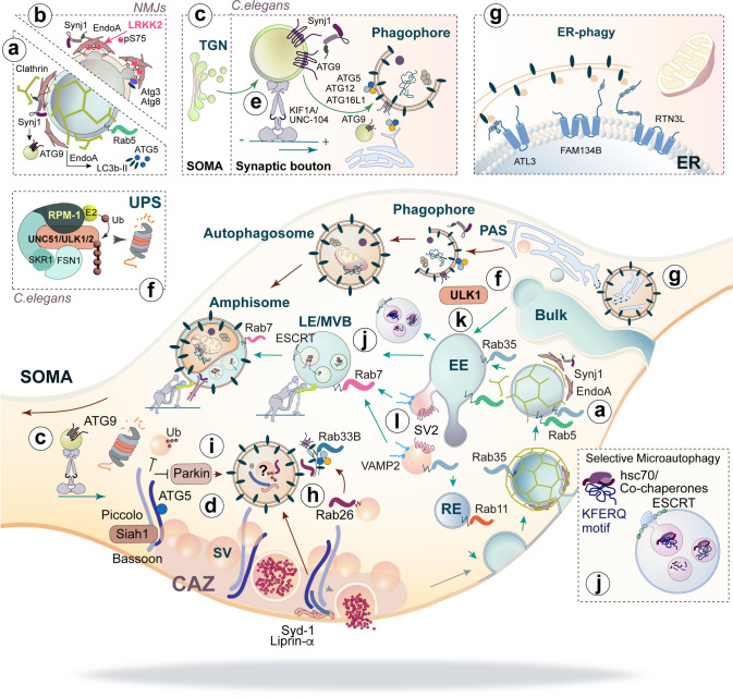
The complex morphology of neurons, the specific requirements of synaptic neurotransmission and the accompanying metabolic demands create a unique challenge for proteostasis. The main machineries for neuronal protein synthesis and degradation are localized in the soma, while synaptic junctions are found at vast distances from the cell body. Sophisticated mechanisms must, therefore, ensure efficient delivery of newly synthesized proteins and removal of faulty proteins. These requirements are exacerbated at presynaptic sites, where the demands for protein turnover are especially high due to synaptic vesicle release and recycling that induces protein damage in an intricate molecular machinery, and where replacement of material is hampered by the extreme length of the axon. In this review, we will discuss the contribution of the two major pathways in place, autophagy and the endolysosomal system, to presynaptic protein turnover and presynaptic function. Although clearly different in their biogenesis, both pathways are characterized by cargo collection and transport into distinct membrane-bound organelles that eventually fuse with lysosomes for cargo degradation. We summarize the available evidence with regard to their degradative function, their regulation by presynaptic machinery and the cargo for each pathway. Finally, we will discuss the interplay of both pathways in neurons and very recent findings that suggest non-canonical functions of degradative organelles in synaptic signalling and plasticity.

Keywords: Autophagy; Axonal boutons; Endolysosomal system; Proteostasis; Synaptic plasticity.


Conflict of interest statement

All authors have read the manuscript and have no competing interests.

Figures

Fig. 1
Fig. 1
Overview of the two pathways that collect and deliver axonal cargo for degradation in lysosomes. Neurons pose a highly compartmentalized morphology endowed with a dendritic tree, the soma and a very long axon (in orange), and are decorated by many synaptic contacts (inset). Synapses are sites with high turnover rates and metabolic demands, where elements of autophagy, endolysosomal system and proteasomal-mediated degradation are present. Two main degradative pathways exist in axons that deliver cargo to lysosomes (described in the lower box) localized in the soma. In autophagy, autophagosome formation is initiated at distal axons at the phagophore assembly site (PAS). Cargo is sequestered by the phagophore which closes generating autophagosomes that are decorated by the autophagy adaptor LC3b-II. Autophagosomes are retrogradely transported in a dynein-dependent manner to the soma, where they fuse with degradative lysosomes generating autolysosomes. The endolysosomal pathway typically receives cargo from the plasma membrane by clathrin-mediated endocytosis and its fate is decided in early-endosomes (EE), organelles abundant in Rab5. Cargo is recycled back to the plasma membrane via recycling endosomes (RE) or is sent for degradation upon internalization into intraluminal vesicles by the ESCRT complex present in late endosomes (LE) and multivesicular bodies (MVB). EE matures into LE/MVB that are Rab7-positive. Cargo is then degraded in endolysosomes that originate from the fusion of LE/MVB with lysosomes. Amphisomes are generated upon the fusion of LE/MVB with autophagosomes and this is a necessary step for the acquisition of retrograde motors by autophagosomes. Ub ubiquitinated receptors
Fig. 2
Fig. 2
Molecular regulators and substrates of autophagy and the endolysosomal systems at presynaptic boutons. Presynaptic proteins regulate autophagy and autophagosome biogenesis at boutons. a, b EndoA and Synj1 mediate clathrin-mediated endocytosis and autophagosome formation. While phosphorylation of EndoA by LRKK2 regulates its insertion into and curvature of membranes (a), the phosphatase activity of the Synj1 SAC1 domain hydrolyses PIPs that are essential for autophagosome formation (b) and both EndoA and Synj1 might control the delivery of pre-autophagosomal membranes to the forming autophagosome via a direct interaction of Synj1 with ATG9 (c). d The active zone protein Bassoon represses local autophagy by sequestering ATG5, whereas autophagy turnover of another two presynaptic proteins, Liprin-α and Syd-1 controls synaptogenesis at Drosophila NMJs. e, f In C. elegans, autophagosome formation is regulated by the kinesin KIF1A/UNC-104 that mediates the anterograde transport of ATG9 (e), and the UPS, which controls the levels of the autophagy initiator UNC-51/ULK1/2 (f). gi Presynaptic organelles are also substrates of autophagy. ER-phage takes place at boutons and several adaptors such as ATL3, FAM134B and RTN3L have been described (g). Degradation of SVs by autophagy is regulated by Rab26, which is involved in their delivery to autophagy membranes labelled by Rab33B (h), and Bassoon which mediates the ubiquitination of SV proteins via Siah and Parkin (i). Entry of integral proteins into the endolysosomal system occurs by clathrin-mediated or -independent endocytosis but proteins carrying a KFERQ motif might also enter this pathway by “endosomal microautophagy” (j). These proteins are recognized by the chaperone Hsc-70 which guides them to the endosomal membrane. Besides degradation by autophagy, SVs also undergo degradation via the endolysosomal system and their endosomal trafficking is regulated by Rab35 which has been shown to recruit ESCRT to SVs (k). SV2 and VAMP2 are degraded via this pathway, while other SV proteins like Synaptotagmin 1 are independent of this pathway (l)
Fig. 3
Fig. 3
Local TrkB signalling at single boutons from TrkB-amphisomes containing SIPA1L2 and its potential implications for presynaptic plasticity. TrkB-amphisomes arise at presynaptic boutons by fusion of autophagosomes and late endosomes (a). Stx17/SNAP29/VAMP8 might control the fusion of both organelles. Myosin VI enable the delivery of endosomes to autophagosomes and its absence leads to an accumulation of immature autophagosomes and a reduced rate of protein aggregate clearance (a, inset). SIPA1L2 mediates the retrograde trafficking of TrkB-amphisomes by directly interacting with Snapin, the adaptor to dynein motors. RapGAP activity of SIPA1L2 is controlled by a direct interaction with the autophagy adaptor LC3b-II that in turns control both, the velocity (b) and signalling properties of the complex in a way that reduced RapGAP activity enables TrkB signalling. PKA activity promotes the stop of the complex at single boutons, reduces SIPA1L2 RapGAP activity that promotes local ERK activation and enhances neurotransmitter release (c). During these synaptic stopovers, TrkB-amphisomes might collect additional cargo, i.e., active TrkB receptors that would enhance the long-range signalling capabilities of the organelle (d). Trafficking of TrkB receptors in amphisomes would benefit from the double-membrane structure of autophagosomes, since TrkB receptors would be incorporated in the outer membrane and, therefore, separated from degradative cargo enclosed within the autophagosome inner membrane (c, d)

Similar articles

Cited by

References

    1. Li XG, Somogyi P, Ylinen A, Buzsaki G. The hippocampal CA3 network: an in vivo intracellular labeling study. J Comp Neurol. 1994;339(2):181–208. doi: 10.1002/cne.903390204. - DOI - PubMed
    1. Sik A, Tamamaki N, Freund TF. Complete axon arborization of a single CA3 pyramidal cell in the rat hippocampus, and its relationship with postsynaptic parvalbumin-containing interneurons. Eur J Neurosci. 1993;5(12):1719–1728. doi: 10.1111/j.1460-9568.1993.tb00239.x. - DOI - PubMed
    1. Rangaraju V, Calloway N, Ryan TA. Activity-driven local ATP synthesis is required for synaptic function. Cell. 2014;156(4):825–835. doi: 10.1016/j.cell.2013.12.042. - DOI - PMC - PubMed
    1. Harris JJ, Jolivet R, Attwell D. Synaptic energy use and supply. Neuron. 2012;75(5):762–777. doi: 10.1016/j.neuron.2012.08.019. - DOI - PubMed
    1. Ziv NE. Maintaining the active zone: demand, supply and disposal of core active zone proteins. Neurosci Res. 2018;127:70–77. doi: 10.1016/j.neures.2017.09.014. - DOI - PubMed

Substances

LinkOut - more resources