. 2021 Apr;162(4):434-451.
doi: 10.1111/imm.13297. Epub 2021 Jan 4.

The human natural anti-αGal antibody targets common pathogens by broad-spectrum polyreactivity

Affiliations

The human natural anti-αGal antibody targets common pathogens by broad-spectrum polyreactivity

Jens Magnus Bernth Jensen et al. Immunology. 2021 Apr.

Abstract

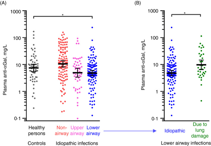
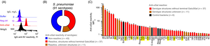
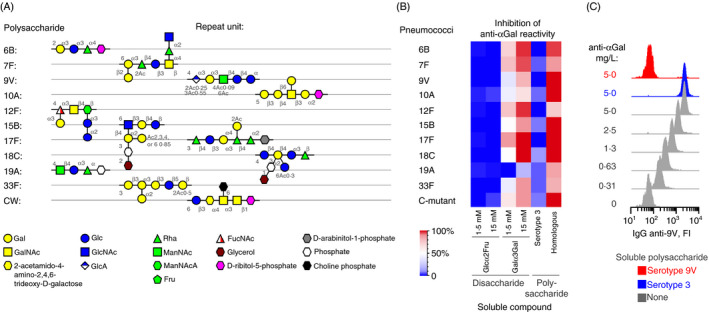
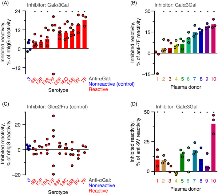
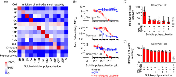
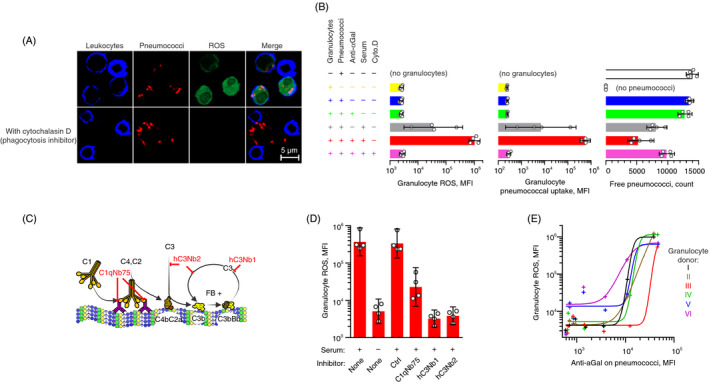
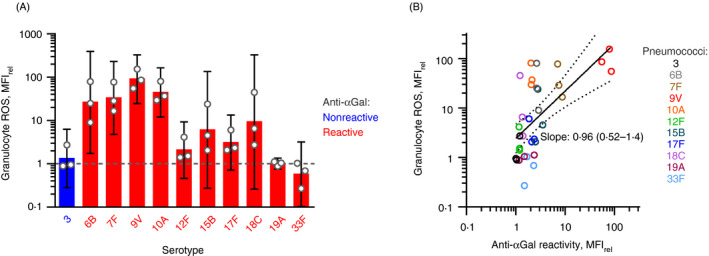
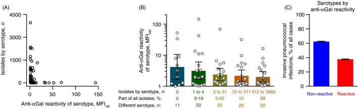
Naturally occurring antibodies are abundant in human plasma, but their importance in the defence against bacterial pathogens is unclear. We studied the role of the most abundant of such antibodies, the antibody against terminal galactose-α-1,3-galactose (anti-αGal), in the protection against pneumococcal infections (Streptococcus pneumonia). All known pneumococcal capsular polysaccharides lack terminal galactose-α-1,3-galactose, yet highly purified human anti-αGal antibody of the IgG class reacted with 48 of 91 pneumococcal serotypes. Anti-αGal was found to contain multiple antibody subsets that possess distinct specificities beyond their general reactivity with terminal galactose-α-1,3-galactose. These subsets in concert targeted a wide range of microbial polysaccharides. We found that anti-αGal constituted up to 40% of the total antibody reactivity to pneumococci in normal human plasma, that anti-αGal drives phagocytosis of pneumococci by human neutrophils and that the anti-αGal level was twofold lower in patients prone to pneumococcal infections compared with controls. Moreover, during a 48-year period in Denmark, the 48 anti-αGal-reactive serotypes caused fewer invasive pneumococcal infections (n = 10 927) than the 43 non-reactive serotypes (n = 18 107), supporting protection on the population level. Our findings explain the broad-spectrum pathogen reactivity of anti-αGal and support that these naturally occurring polyreactive antibodies contribute significantly to human protective immunity.

Keywords: antibodies; epitopes; flow cytometry; microbiota.


Conflict of interest statement

The authors declare no competing financial and commercial interests in relation to the manuscript.

Figures

FIGURE 1
FIGURE 1
Low plasma levels of anti‐αGal antibody in humans with increased burden of lower airway infections. Anti‐αGal antibody was quantified in plasma samples by solid‐phase immunoassay. (A) Comparison of anti‐αGal levels in healthy persons (control, n = 60) and in patients suffering from recurrent infections (n = 289) to a degree prompting experienced medical specialist to suspect causative primary immunodeficiency. Patients were categorized according to their dominating site of infection: (i) non‐airways (n = 118), (ii) upper airways (n = 53) and (iii) lower airways (n = 118). The black bars show the geometric means with 95% confidence intervals. The control group was compared with each of the patient subgroups based on bootstrap sampling distributions (Figure S2), and significant group difference (P < 0·05) in the anti‐αGal levels is marked by a grey horizontal line and asterisk. (B) As in panel A but for patients with idiopathic lower‐airway infections (from panel A) compared with an additional control group comprised of patients with severe lung damage (lung transplantation candidates) and thus highly increased tendency to acquire lower‐airway infections (n = 34)
FIGURE 2
FIGURE 2
The anti‐αGal antibody binds most pneumococcal serotypes. Flow cytometry analyses showing antibody reactivity with various bacteria. (A) Example plot showing antibody reactivity with serotype 9V pneumococci after incubation with buffer only, negative control IgG (IgG anti‐CD20, i.e. irrelevant specificity) at 10 mg/L, purified human anti‐αGal at 5 mg/L or normal human IgG (nhIgG) at 500 mg/L. NhIgG is expected to contain antibodies to pneumococci and served as positive control. IgG on the pneumococci was assayed with fluorescently labelled rabbit F(ab’)2 anti‐human IgG. ‘FI’: Fluorescence intensity. (B) Pie‐chart summarizing reactivity of anti‐αGal (5 mg/L) with 91 serotypes of S. pneumoniae. (C) Level of reactivity with the reactive pneumococcal serotypes and five control bacterial strains. Control strains were E. coli O86 (EcO86) and four unencapsulated pneumococcal strains: C‐mutant (covered by a thick layer of cell wall (CW) polysaccharide), Rough1, Rough2 and Rough4. The columns are the mean reactivity (MFIrel) and standard deviation of two separate experiments. MFIrel was the MFI in experiments with anti‐αGal relative to the MFI in the same experiment but without primary antibody (fluorescently labelled anti‐hIgG was present in both experiments). A positive antibody reaction was defined as a mean MFIrel at least two standard deviations above 1·10 (corresponding to the highest reactivity observed in parallel and equal experiments but with the negative control IgG anti‐CD20 as primary antibody). The figure shows that anti‐αGal reacts with numerous serotypes although they do not possess terminal Galα3Gal in their polysaccharide structure (red columns) (see also Figure 3). Several serotypes demonstrate higher reactivity than the positive control strain, EcO86. The serotypes highlighted in yellow were selected for further experiments
FIGURE 3
FIGURE 3
The anti‐αGal antibody is polyreactive and binds distinct capsular polysaccharides. (A) Schematic structures of the repeat unit in selected pneumococcal polysaccharides (capsular polysaccharide serotypes and CW polysaccharide). (B) Heatmap showing the inhibitory effect of various soluble compounds on the reaction between anti‐αGal and each of 10 pneumococcal serotypes and the C‐mutant strain. The concentration of the used anti‐αGal was 5 mg/L. For each strain, inhibition by the following soluble compounds was tested: Glcα2Fru (negative control), Galα3Gal, capsular polysaccharide of serotype 3 (negative control, intact pneumococci of serotype 3 did not react with anti‐αGal), and the homologous polysaccharide (i.e. capsular polysaccharide of the same serotype as the examined strain and CW polysaccharide for the C‐mutant strain). The polysaccharide concentration was 125 mg/L. The level of inhibition was determined by flow cytometry as the mean of two separate experiments. (C) Example plots of raw data sampled for serotype 9V pneumococci for quantification of anti‐αGal reactivity and inhibition by homologous serotype 9V polysaccharide (red) and heterologous serotype 3 polysaccharide (blue). The reactivity was calculated by means of standard curves as exemplified in Figure S7
FIGURE 4
FIGURE 4
The anti‐αGal antibody comprises significant parts of human anti‐pneumococcal antibody reactivities. Inhibition of IgG reactivity with different serotypes of pneumococci by the presence of different disaccharides. Reactivity was quantified by flow cytometry. (A) Normal human IgG (nhIgG) was used as source of anti‐pneumococcal IgG. The reactivity with each of ten serotypes was challenged by Galα3Gal at 37 mM. Circles show the results of separate experiments, and the bars are the geometric mean for reactivity with each serotype (note that geometric means are not defined for datasets including negative values). Results are marked by an asterisk when the 95% confidence interval of the mean does not contain 0 (and therefore is considered significantly different from 0). The inhibited antibodies reacted with the soluble Galα3Gal and are therefore anti‐αGal. Thus, the level of lost reactivity represents the percentage of anti‐αGal that comprised the anti‐pneumococcal reactivity of nhIgG. (B) As in the previous panel, except that the reactions were challenged by Glcα2Fru. Glcα2Fru did not inhibit the reactivity of nhIgG with any serotype. (C) As in panel A, except that the antibody source was plasma samples from different healthy persons tested against serotype 7F pneumococci. (D) As in the previous panel, except serotype 9V pneumococci were used as target pneumococci
FIGURE 5
FIGURE 5
The anti‐αGal antibody contains antibody subsets of distinct polyreactivity. Flow cytometry analyses of anti‐αGal reactivity with target cells in the presence of soluble polysaccharides. (A) Heatmap showing results for various cells. Anti‐αGal was added at 5 mg/L and polysaccharides at 125 mg/L. Target cells were ten different serotypes of pneumococci, C‐mutant, EcO86 and pig erythrocytes (carries surface terminal Galα3Gal). The level of inhibition was determined as the mean of two separate experiments. The left downward diagonal of red squares shows that the homologous polysaccharide inhibited reactivity better than the heterologous polysaccharides. (B) Reactivity of anti‐αGal with each of three encapsulated pneumococcal strains in the presence of increasing concentration of either CW polysaccharide or homologous capsular polysaccharide. Anti‐αGal was added at 2·0 mg/L (serotypes 9V and 12F) or 10 mg/L (serotype 15B). Mean and standard deviation of two separate experiments. Centre (serotype 12F): vertical arrows indicate half‐maximal inhibitory concentration of each polysaccharide. The panel shows that CW polysaccharide was a more potent inhibitor than the homologous capsular polysaccharide for this serotype 12F strain. Also, neither substance alone caused complete inhibition. Bottom (serotype 15B): CW polysaccharide inhibited at most half of the reactivity, indicating that only a subset of the anti‐αGal antibodies that reacted with this strain possessed specificity for CW polysaccharide. (C) Residual reactivity of anti‐αGal at 2·0 mg/L with the strain of serotype 12F in the presence of soluble polysaccharides, each added at 125 mg/L. Mean with 95% confidence intervals of three separate experiments. (D) As in the previous panel, but with the strain of serotype 15B as target cells and anti‐αGal at 10 mg/L
FIGURE 6
FIGURE 6
Polyreactive anti‐αGal antibodies drive phagocytosis of serotype 9V pneumococci in a complement‐dependent manner. Fluorescently labelled serotype 9V pneumococci were opsonized in 10% human hypogammaglobulinaemia serum (‘serum’) supplemented with anti‐αGal at 20 mg/L (unless otherwise is stated) before feeding to plasma‐depleted human blood cells labelled with dihydrorhodamine‐123. Dihydrorhodamine‐123 (non‐fluorescent) is oxidized to rhodamine‐123 (fluorescent) by intra‐phagosomal reactive oxygen species (ROS) produced in phagocytosing granulocytes. Confocal microscopy in A and flow cytometry data in B, D and E. (A) Experiments with and without the phagocytosis inhibitor cytochalasin D. The panel shows that pneumococci and ROS were inside the leucocytes when the phagocytosis inhibitor was omitted, whereas no ROS or pneumococci were inside the leucocytes when the phagocytosis inhibitor was present. (B) Effect of constituents in the experiments on granulocyte ROS (left), pneumococcal uptake (centre) and clearance of free pneumococci from the supernatant (right). Bars represent mean with 95% confidence intervals for different leucocyte donors (n = 4). (C) Illustration of complement activation by IgG antibodies bound on antigen and the points of action for the specific complement inhibitors used in the experiments shown in the following panel. ‘C1qNb75’, blocks C1q binding to immunoglobulin; ‘hC3Nb1’, blocks C3 cleavage by alternative pathway; and ‘hC3Nb2’, blocks C3 cleavage by classical and alternative pathways. (D) Granulocyte ROS induced by pneumococci, opsonized with anti‐αGal at 5 mg/L in the presence of inhibitors of selective complement factors. ‘Ctrl’, nanobody of irrelevant specificity. Mean with 95% confidence intervals for different leucocyte donors (n = 4). (E) Granulocyte ROS as function of the density of anti‐αGal on opsonized pneumococci. For each of six leucocyte donors, experiments were performed with the following concentrations of anti‐αGal: 0, 0·020, 0·078, 0·31, 1·3, 5 and 20 mg/L. Curves were fitted in sigmoid models (R 2 ≥ 0·92)
FIGURE 7
FIGURE 7
Binding of polyreactive anti‐αGal antibodies in general conveys phagocytosis of pneumococcal serotypes. (A) The opsonic effect of anti‐αGal for each 11 different pneumococcal serotypes. Flow cytometry data (MFIrel) for opsonization with hypogammaglobulinaemia serum (‘serum’) with added anti‐αGal compared with opsonization with hypogammaglobulinaemia serum only. Serum concentration was 10%. Concentration of added anti‐αGal was 20 mg/L. Mean with 95% confidence intervals for three leucocyte donors. (B) The general opsonic effect of anti‐αGal for reactive pneumococci. Granulocyte ROS (from previous panel, on y‐axis) as function of antibody binding on pneumococci after opsonization with anti‐αGal and hypogammaglobulinaemia serum relative to hypogammaglobulinaemia serum alone. The regression curve with 95% confidence interval, determined on log10–log10 scale, is shown together with the point estimate of the curve´s slope (with 95% confidence interval). For these serotypes, the data show a positive correlation between the level of anti‐αGal reactivity and the phagocytosis signal
FIGURE 8
FIGURE 8
Relationship between anti‐αGal reactivity of pneumococcal serotypes and their occurrence among invasive infections in humans. All recorded cases of invasive infections due to encapsulated pneumococci in Denmark between 1966 and 2014 (n = 29 034) were included. (A) The number of isolates of each serotype is shown as a function of the anti‐αGal reactivity of that serotype (as determined in the present study). (B) Anti‐αGal reactivity for serotypes grouped according to the number of recorded isolates. For each group, the accumulated contribution to invasive pneumococcal infections and number of different serotypes included in the group are shown. Geometric mean with 95% confidence intervals. Anti‐αGal reactivity tended to be lower for the more prevalent serotypes. (C) The percentage of invasive pneumococcal infections caused by serotypes grouped according to their anti‐αGal reactivity. Error bars are 95% confidence interval. The non‐reactive serotypes were more prevalent than the reactive serotypes

Similar articles

Cited by

References

    1. Springer GF, Williamson P, Brandes WC. Blood group activity of gram‐negative bacteria. J Exp Med. 1961; 113:1077–93. - PMC - PubMed
    1. Galili U, Rachmilewitz EA, Peleg A, Flechner I. A unique natural human IgG antibody with anti‐alpha‐galactosyl specificity. J Exp Med. 1984; 160:1519–31. - PMC - PubMed
    1. Tangvoranuntakul P, Gagneux P, Diaz S, Bardor M, Varki N, Varki A, et al. Human uptake and incorporation of an immunogenic nonhuman dietary sialic acid. Proc Natl Acad Sci USA. 2003; 100:12045–50. - PMC - PubMed
    1. Galili U. Anti‐Gal: an abundant human natural antibody of multiple pathogeneses and clinical benefits. Immunology. 2013; 140:1–11. - PMC - PubMed
    1. Galili U, Clark MR, Shohet SB, Buehler J, Macher BA. Evolutionary relationship between the natural anti‐Gal antibody and the Gal alpha 1–3Gal epitope in primates. Proc Natl Acad Sci USA. 1987; 84:1369–73. - PMC - PubMed

Publication types

LinkOut - more resources