Review
. 2021 Jan;20(1):68-80.
doi: 10.1016/S1474-4422(20)30412-9.

APOE and Alzheimer's disease: advances in genetics, pathophysiology, and therapeutic approaches

Affiliations
Review

APOE and Alzheimer's disease: advances in genetics, pathophysiology, and therapeutic approaches

Alberto Serrano-Pozo et al. Lancet Neurol. 2021 Jan.

Erratum in

  • Correction to Lancet Neurol 2021; 20: 68-80.
    [No authors listed] [No authors listed] Lancet Neurol. 2021 Feb;20(2):e2. doi: 10.1016/S1474-4422(21)00004-1. Epub 2021 Jan 20. Lancet Neurol. 2021. PMID: 33484655 No abstract available.

Abstract

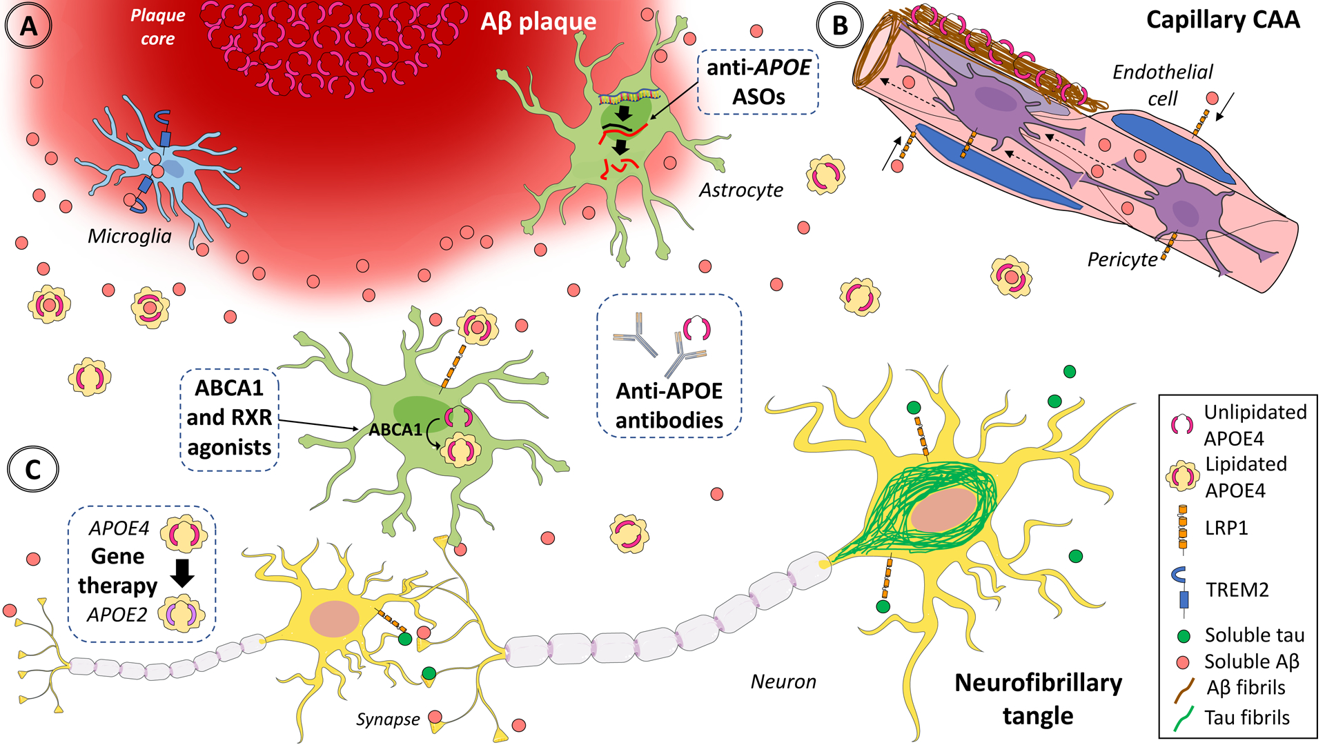
The APOE ε4 allele remains the strongest genetic risk factor for sporadic Alzheimer's disease and the APOE ε2 allele the strongest genetic protective factor after multiple large scale genome-wide association studies and genome-wide association meta-analyses. However, no therapies directed at APOE are currently available. Although initial studies causally linked APOE with amyloid-β peptide aggregation and clearance, over the past 5 years our understanding of APOE pathogenesis has expanded beyond amyloid-β peptide-centric mechanisms to tau neurofibrillary degeneration, microglia and astrocyte responses, and blood-brain barrier disruption. Because all these pathological processes can potentially contribute to cognitive impairment, it is important to use this new knowledge to develop therapies directed at APOE. Several therapeutic approaches have been successful in mouse models expressing human APOE alleles, including increasing or reducing APOE levels, enhancing its lipidation, blocking the interactions between APOE and amyloid-β peptide, and genetically switching APOE4 to APOE3 or APOE2 isoforms, but translation to human clinical trials has proven challenging.


Figures

Figure
Figure. Multifaceted effects of APOE in the brain and potential strategies to decrease APOE4 and increase APOE2 levels.
In the healthy brain, APOE is expressed and secreted predominantly by astrocytes, and to a lesser extent by microglia. Most brain APOE is lipidated by the ATP-binding cassettes A1 (ABCA1) and G1 (ABCG1) and lipidated APOE is internalized via APOE receptors such low-density lipoprotein receptor-related protein 1 (LRP1), which is expressed in astrocytes, neurons, vascular smooth muscle cells, endothelial cells, and pericytes. In the Alzheimer’s disease brain, astrocytes and microglia react to (A) dense-core Aβ plaques, (B) cerebral amyloid angiopathy-laden arteries and capillaries, and (C) neurofibrillary tangles, activating transcriptional programmes that include APOE mRNA up-regulation in microglia, and down-regulation in astrocytes, and lead to altered lipid metabolism (not shown). APOE directly interacts with both soluble and fibrillar Aβ. Relative to APOE3 and APOE2, APOE4 promotes Aβ seeding and aggregation in oligomers and fibrils and reduces its clearance from the interstitial fluid, potentially leading to Aβ deposition as dense-core (Thioflavin-S positive) amyloid plaques and cerebral amyloid angiopathy together with APOE. This evidence suggests that decreasing APOE (especially APOE4) expression or blocking the effects of APOE4 or enhancing the effects APOE2 would be beneficial (dashed boxes). Experimental approaches to achieving these outcomes include lowering APOE4 levels with isoform-specific antisense oligonucleotides or antibodies, which could also target lipid-poor APOE associated with plaques. Alternatively, APOE4 could be switched to APOE3 or APOE2, or APOE2 could be added,, with gene therapy. Last, APOE lipidation could be enhanced with RXR and ABCA1 or ABCG1 agonists to improve APOE4 receptor-mediated internalization and lower Aβ in the interstitial fluid. Dashed boxes illustrate the most promising therapeutic approaches. ASO=antisense oligonucleotides. Aβ=amyloid-β peptide. CAA=cerebral amyloid angiopathy. TREM2=triggering receptor expressed in myeloid cells 2. RXR=retinoid X receptor. Adapted from Servier Medical Art by Servier (https://smart.servier.com/category/medical-specialties/neurology/), which is licensed under a Creative Commons Attribution 3.0 Unported License (CC BY 3.0).

References

    1. Kunkle BW, Grenier-Boley B, Sims R, et al. Genetic meta-analysis of diagnosed Alzheimer’s disease identifies new risk loci and implicates Aβ, tau, immunity and lipid processing. Nat Genet 2019; 51: 414–30. - PMC - PubMed
    1. Reiman EM, Arboleda-Velasquez JF, Quiroz YT, et al. Exceptionally low likelihood of Alzheimer’s dementia in APOE2 homozygotes from a 5,000-person neuropathological study. Nat Commun 2020; 11: 667. - PMC - PubMed
    1. Arboleda-Velasquez JF, Lopera F, O’Hare M, et al. Resistance to autosomal dominant Alzheimer’s disease in an APOE3 Christchurch homozygote: a case report. Nat Med 2019; 25: 1680–3. - PMC - PubMed
    1. Belloy ME, Napolioni V, Han SS, Le Guen Y, Greicius MD, Alzheimer’s Disease Neuroimaging Initiative. Association of Klotho-VS Heterozygosity With Risk of Alzheimer Disease in Individuals Who Carry APOE4. JAMA Neurol 2020; published online April 13. DOI:10.1001/jamaneurol.2020.0414. - DOI - PMC - PubMed
    1. Zhou X, Chen Y, Mok KY, et al. Non-coding variability at the APOE locus contributes to the Alzheimer’s risk. Nat Commun 2019; 10: 3310. - PMC - PubMed