Comparative Study
. 2021 Apr;10(4):598-609.
doi: 10.1002/sctm.20-0234. Epub 2020 Dec 19.

Comparison of concentrated fresh mononuclear cells and cultured mesenchymal stem cells from bone marrow for bone regeneration

Affiliations
Comparative Study

Comparison of concentrated fresh mononuclear cells and cultured mesenchymal stem cells from bone marrow for bone regeneration

Fengzhou Du et al. Stem Cells Transl Med. 2021 Apr.

Abstract

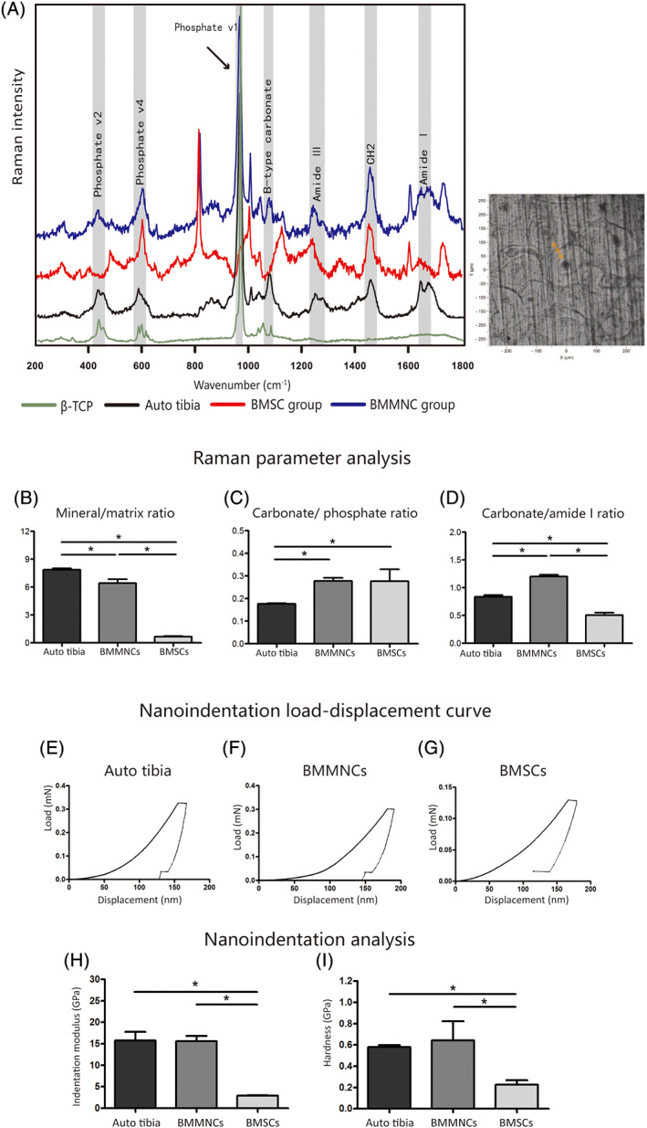
Autologous bone marrow mononuclear cell (BMMNC) transplantation has been widely studied in recent years. The fresh cell cocktail in BMMNCs, without going through the in vitro culture process, helps to establish a stable microenvironment for osteogenesis, and each cell type may play a unique role in bone regeneration. Our study compared the efficacy of concentrated fresh BMMNCs and cultured bone marrow-derived mesenchymal stem cells (BMSCs) in Beagle dogs for the first time. Fifteen-millimeter segmental bone defects were created in the animals' tibia bones. In BMMNCs group, the defects were repaired with concentrated fresh BMMNCs combined with β-TCP (n = 5); in cultured BMSC group, with in vitro cultured and osteo-induced BMSCs combined with β-TCP (n = 5); in scaffold-only group, with a β-TCP graft alone (n = 5); and in blank group, nothing was grafted (n = 3). The healing process was monitored by X-rays and single photon emission computed tomography. The animals were sacrificed 12 months after surgery and their tibias were harvested and analyzed by microcomputed tomography and hard tissue histology. Moreover, the microstructure, chemical components, and microbiomechanical properties of the regenerated bone tissue were explored by multiphoton microscopy, Raman spectroscopy and nanoindentation. The results showed that BMMNCs group promoted much more bone regeneration than cultured BMSC group. The grafts in BMMNCs group were better mineralized, and they had collagen arrangement and microbiomechanical properties similar to the contralateral native tibia bone. These results indicate that concentrated fresh bone marrow mononuclear cells may be superior to in vitro expanded stem cells in segmental bone defect repair.

Keywords: bone marrow-derived mesenchymal stem cells; bone regeneration; concentrated bone marrow mononuclear cells; β-TCP.


Conflict of interest statement

The authors declared no potential conflicts of interests.

Figures

FIGURE 1
FIGURE 1
Bone marrow mononuclear cells (BMMNCs)/β‐TCP repaired segmental defects in Beagle dogs. A‐D, Representative X‐ray images showing the healing process of a subject in the BMMNC group (n = 5) at 14 days (A), 3 months (B), 6 months (C), and 12 months (D). E‐H, Representative X‐ray images of a subject in the bone marrow‐derived mesenchymal stem cell (BMSC) group (n = 5) at 14 days (E), 3 months (F), 6 months (G), and 12 months (H). I‐L, SPECT and SPECT/CT fusion images showing the concentration in the graft area in the BMMNC group (I, J) and the BMSC group (K, L). M, Comparison of the uptake ratio in the SPECT of the two groups (t test)
FIGURE 2
FIGURE 2
Gross observations and micro‐CT analysis showing massive bone regeneration after bone marrow mononuclear cells (BMMNCs)/β‐TCP grafting. A‐F, Gross view, section view, and micro‐CT images of the tibia at 12 months after surgery in the BMMNC (A‐C) and the bone marrow‐derived mesenchymal stem cell (BMSC) (D‐F) groups. The section views are displayed in proximal, distal and transverse segments. G‐I, Micro‐CT analysis results. BV (G) and BV/TV (H) were significantly higher in the BMMNC group. TMD (I) in the BMMNC and BMSC groups were not significantly different but were lower than in the contralateral autotibia (t test)
FIGURE 3
FIGURE 3
Histology of the grafts in the bone marrow mononuclear cell (BMMNC) and bone marrow‐derived mesenchymal stem cell (BMSC) groups at 12 months after surgery. A‐E, Methylene blue‐acid fuchsine staining in the BMMNCs/β‐TCP treated animals. Cross and longitudinal sections show newly formed bone tissue (bright pink) and an undegraded scaffold (A). The macropores of the scaffold were filled with bone tissue (B). Blood vessels were located in the Haversian canals (C) and on the walls of the macropores. The Harversian system could also be visualized in the regenerated bone tissue around the scaffold (D, E). F‐J, Modified Goldner's trichrome staining of the graft from the BMMNC group. Newly formed bone around the scaffold included hypomineralized bone tissue (red) (F), which was located at the center of the osteons (I, J). The bone tissue in the macro pores was well‐mineralized (blue) (G), and the osteocytes were well‐defined between the lamellae (H). K‐T, Histology of the animals in the BMSC group. Newly formed bone tissue was visualized inside the scaffold (K‐M), but the pores near the center of the graft were filled with soft tissues (blue) (N, O). There was a clear interface with the native cortical bone (P). The regenerated bone tissue was not fully mineralized in most areas (orange) (S, T)
FIGURE 4
FIGURE 4
Microstructure of the regenerated bone tissue investigated by multiphoton microscopy. A‐F, Bone marrow mononuclear cell (BMMNC) group. Two‐photon excitation fluorescence (TPEF) image showing the bone matrix, blood vessels, and osteocytes in the macro pores of the scaffold (A). The collagen fibers visualized by SHG were arranged concentrically or aligned in layers near the scaffold (B). The merged image showing blood vessels in the osteons (C). Regenerated bone outside the scaffold showed a deep lacunas (D) and well‐organized collagens (E, F). G–I, Bone marrow‐derived mesenchymal stem cell (BMSC) group. No lacunas were observed (G), and the collagen fibers were disorganized (H, I). J‐L, Contralateral auto tibia. TPEF and SHG images showing well‐formed lacunas (J) and osteons (K, L)
FIGURE 5
FIGURE 5
Bone compositional and biomechanical analysis. A, Left, typical spectra of bone tissue from the bone marrow mononuclear cell (BMMNC) group, the bone marrow‐derived mesenchymal stem cell (BMSC) group, the autotibia and β‐TCP scaffold. Right, the test points randomly chosen along the radii of the osteons (B‐D) Raman parameter analysis (n = 3 auto tibia, n = 4 BMMNCs, n = 3 BMSCs) of the mineral/matrix ratio (B), carbonate/phosphate ratio (C), and carbonate/amide I ratio (D) (t test). F‐H, Typical nanoindentation load‐displacement curve of specimens from the contralateral autotibia (F), the BMMNC group (G), and the BMSC group (H). I,J, Nanoindentation analysis (n = 3 auto tibia, n = 4 BMMNCs, and n = 3 BMSCs) of the indentation modulus (I) and hardness (J) (t test)
FIGURE 6
FIGURE 6
Analysis of cytokines in the bone marrow mononuclear cell (BMMNC) suspension. A‐E, Relative levels of IL‐2 (E), IL‐6 (F), IL‐8 (G), RAGE (H), and SCF (I) in PBP (n = 3), BMA (n = 3), and BMMNCs (n = 10) of the Beagle dogs (t test)

Similar articles

Cited by

References

    1. Wise JK, Alford AI, Goldstein SA, Stegemann JP. Comparison of uncultured marrow mononuclear cells and culture‐expanded mesenchymal stem cells in 3D collagen‐chitosan microbeads for orthopedic tissue engineering. Tissue Eng Part A. 2014;20(1–2):210‐224. - PMC - PubMed
    1. Lekholm U, Wannfors K, Isaksson S, Adielsson B. Oral implants in combination with bone grafts. A 3‐year retrospective multicenter study using the Branemark implant system. Int J Oral Maxillofac Surg. 1999;28(3):181‐187. - PubMed
    1. Hernigou P, Poignard A, Beaujean F, Rouard H. Percutaneous autologous bone‐marrow grafting for nonunions. Influence of the number and concentration of progenitor cells. J Bone Joint Surg Am. 2005;87(7):1430‐1437. - PubMed
    1. Daltro GC, Fortuna V, de Souza ES, et al. Efficacy of autologous stem cell‐based therapy for osteonecrosis of the femoral head in sickle cell disease: a five‐year follow‐up study. Stem Cell Res Ther. 2015;6:110. - PMC - PubMed
    1. Lee DH, Ryu KJ, Kim JW, Kang KC, Choi YR. Bone marrow aspirate concentrate and platelet‐rich plasma enhanced bone healing in distraction osteogenesis of the tibia. Clin Orthop Relat Res. 2014;472(12):3789‐3797. - PMC - PubMed

Publication types

Substances