. 2021 Jan:63:103167.
doi: 10.1016/j.ebiom.2020.103167. Epub 2020 Dec 18.

Dolutegravir in pregnant mice is associated with increased rates of fetal defects at therapeutic but not at supratherapeutic levels

Affiliations

Dolutegravir in pregnant mice is associated with increased rates of fetal defects at therapeutic but not at supratherapeutic levels

Haneesha Mohan et al. EBioMedicine. 2021 Jan.

Abstract

Background: Dolutegravir (DTG) is a preferred regimen for all people with HIV including pregnant women, but its effects on the fetus are not fully understood. Periconceptional exposure to DTG has been associated with increased rates of neural tube defects (NTDs), although it is unknown whether this is a causal relationship. This has led to uncertainty around the use of DTG in women of reproductive potential.

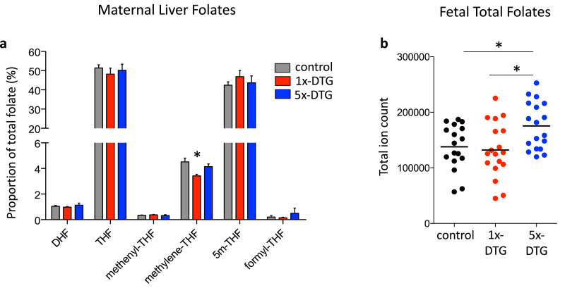
Methods: Pregnant C57BL/6J mice were randomly allocated to control (water), 1x-DTG (2.5 mg/kg-peak plasma concentration ~3000 ng/ml - therapeutic level), or 5x-DTG (12.5 mg/kg-peak plasma concentration ~12,000 ng/ml - supratherapeutic level), once daily from gestational day 0.5 until sacrifice. DTG was administered with 50 mg/kg tenofovir+33.3 mg/kg emtricitabine. Fetal phenotypes were determined, and maternal and fetal folate levels were quantified by mass-spectrometry.

Findings: 352 litters (91 control, 150 1x-DTG, 111 5x-DTG) yielding 2776 fetuses (747 control, 1174 1x-DTG, 855 5x-DTG) were assessed. Litter size and viability rates were similar between groups. Fetal and placenta weights were lower in the 1x-DTG vs. control. Placental weight was higher in the 5x-DTG vs. control. Five NTDs were observed, all in the 1x-DTG group. Fetal defects, including microphthalmia, severe edema, and vascular/bleeding defects were more frequent in the 1x-DTG group. In contrast, defect rates in the 5x-DTG were similar to control. Fetal folate levels were similar between control and 1x-DTG, but were significantly higher in the 5x-DTG group.

Interpretation: Our findings support a causal relationship of DTG at therapeutic doses with increased risk for fetal defects, including NTDs at a rate that is similar that reported in the Tsepamo study for women exposed to DTG-based ART from conception. The non-monotonic dose-response relationship between DTG and fetal anomalies could explain the previous lack of fetal toxicity findings from pre-clinical DTG studies. The fetal folate levels suggest that DTG is unlikely to be an inhibitor of folate uptake.

Funding: This project has been funded with Federal funds from the Eunice Kennedy Shriver National Institute of Child Health and Human Development, National Institutes of Health, Department of Health and Human Services, under Contract No. HHSN275201800001I.


Conflict of interest statement

Declaration of Competing Interest The authors have no competing interests relating to this study. AJC acts as consultant for ViiV Healthcare Limited, with any fees going to support his research program. LS received personal support for participating in a ViiV organized Think Tank.

Figures

Fig 1
Fig. 1
Comparison of pregnancy outcomes between treatment arms. Pregnant mice were treated with 1x-DTG (2.5 mg/kg DTG + TDF/FTC), 5x-DTG (12.5 mg/kg DTG + TDF/FTC), or water as a control starting on gestational day (GD) 0.5 until sacrifice on GD15.5. Schematic representation of the pregnancy mouse model is shown in (a), viability rate in (b), litter average fetal weight in (c), litter average placental weight in (d), and percent increase in maternal weight in (e). Data are shown as dot plots with the line indicating the median. Statistical comparison by Kruskal–Wallis with Dunn's post-test vs control. * p<0.05, ** p<0.01, *** p<0.001. N=90 for control, N=143 for 1x-DTG, and N=100 for 5x-DTG.
Fig 2
Fig. 2
Neural tube defects in fetuses in the 1x-DTG treatment arm. Images of a representative control (a), and the five fetuses with neural tube defects (b–f). Scale bars = 1 mm. (b) Non-viable fetus exhibiting exencephaly, kinky tail, hemorrhage, and growth retardation. (c) Non-viable, edematous fetus exhibiting exencephaly, holoprosencephaly, mandibular aplasia, maxillary prognathism, and bilateral anophthalmia. (d) Non-viable fetus with open forebrain, and growth retardation. (e–f) Viable fetuses exhibiting spina bifida. The region of defect is highlighted by a dashed red box, with a higher magnification of the affected region in the lower images. An H&E section shows evidence of spina bifida in (e) bottom panel. (g) Mean litter rate with 95% confidence interval for neural tube defects in the control (ctr, n=91 litters), 1x-DTG (2.5 mg/kg DTG + TDF/FTC, n=150 litters) and 5x-DTG (12.5 mg/kg DTG + TDF/FTC, n=111 litters). Neural tube defects mean litter rate is significantly higher in the 1x-DTG group vs. control (p<0.05). Statistical comparison by Kruskal–Wallis with Dunn's post-test.
Fig 3
Fig. 3
Eye defects are more common in fetuses in the 1x-DTG treatment arm. Gross morphology of representative fetuses showing normal eyes (a), anophthalmia (b), microphthalmia (c), and coloboma (d). Arrows point to the eye. Scale bars = 1 mm. (e) Mean litter rate with 95% confidence interval for microphthalmia in the control (ctr), 1x-DTG (2.5 mg/kg DTG + TDF/FTC) and 5x-DTG (12.5 mg/kg DTG + TDF/FTC). Mean litter rate for microphthalmia is significantly higher in the 1x-DTG group vs. control (p<0.01). Statistical comparison by Kruskal–Wallis with Dunn's post-test. N=91 litters for control, N=150 litters for 1x-DTG, N=111 litters for 5x-DTG.
Fig 4
Fig. 4
Severe edema is more common in fetuses in the 1x-DTG treatment arm and coincides with vascular leak and lower fetal liver volumes. Gross morphology of representative fetuses, normal/control (a), and severe edema (b–c). Scale bars = 1 mm. Black arrows highlight regions of edema (b, c). (d) H&E stained sections from a control fetus (top) and a 1x-DTG fetus (bottom). Arrows in the left panels point to the area of edema in a 1x-DTG fetus (bottom-left panel) and equivalent area in the control (upper-left panel). Arrows in close ups in the right panels point to areas of blood pooling in the 1x-DTG (lower-right panel) and equivalent area in the control (upper-right panel). Vascular leakage is evidenced by presence of erythrocytes within the edematous area in a 1x-DTG fetus (bottom-right panel). Scale bars = 1 mm and 100 µm in close ups. (e) Mean litter rate with 95% confidence interval for severe edema in the control (ctr), 1x-DTG (2.5 mg/kg DTG + TDF/FTC) and 5x-DTG (12.5 mg/kg DTG + TDF/FTC). Mean litter rate for severe edema is significantly higher in the 1x-DTG group vs. control and 5x-DTG (p<0.01). Statistical comparison by Kruskal–Wallis with Dunn's post-test. N=91 litters for control, N=150 litters for 1x-DTG, N=111 litters for 5x-DTG. (f) Mid-sagittal sections of fetal images acquired by magnetic resonance imaging of a control fetus (top panel) and a 1x-DTG fetus with severe edema (bottom panel). White arrows point to the liver. Scale bars = 1 mm. (g) Fetal liver volume (normalized to fetal weight) is lower in 1x-DTG fetuses with severe edema. Data shown as dot plots with mean and 95% confidence interval. Statistical comparisons by Student's t-test. N=8 fetuses/group from 8 different litters. ** p<0.01.
Fig 5
Fig. 5
Vascular defects are more common in fetuses in the 1x-DTG treatment arm. Gross morphology of representative fetuses showing petechiae (b), cranial bleed (c), spinal bleed (d), and hemorrhagic bleed (e), compared with a normal fetus (a). Scale bars = 1 mm. Mean litter rate with 95% confidence interval for petechiae in (f) and cranial or spinal bleeds in (g) in the control (ctr), 1x-DTG (2.5 mg/kg DTG + TDF/FTC) and 5x-DTG (12.5 mg/kg DTG + TDF/FTC). Both types of vascular disorder are significantly more prevalent in the 1X-DTG group vs. control (p<0.01 for petechial, p<0.001 for cranial/spinal bleed). Statistical comparison by Kruskal–Wallis with Dunn's post-test. N=91 litters for control, N=150 litters for 1x-DTG, N=111 litters for 5x-DTG. (h–i) H&E sections from control and 1x-DTG fetuses. (h) Intra-peritoneal hemorrhage in a 1x-DTG fetus, which also had a cranial bleed (as seen in C). (i) Pericardial hemorrhage in a 1x-DTG fetus which also had a spinal bleed (as seen in D). Close ups are shown in the right panels. Scale bar = 1 mm and 0.5 mm in close ups.
Fig 6
Fig. 6
Maternal liver folate profiles and fetal total folate levels differ between treatment arms. Folate profile in maternal liver (a) and total folate levels in whole fetuses (b) collected on gestational day 11.5 and assessed by mass spectrometry. Statistical comparisons by one-way ANOVA with Bonferroni's multiple comparisons post-test. Maternal folate profiles differ only in a reduced methylene-THF proportion in the 1x-DTG group vs. control. Fetal total folates are significantly higher in the 5x-DTG group vs. control (* p<0.05 vs. control and 1x-DTG). For maternal folates N=6/group. For fetal folates N=18 fetuses/group from 6 different litters. Data are shown as means with SEM in (a) and as dots plots with the line indicating the mean in (b). DHF, dihydropholate; THF, tetrahydrofolate; 5m-THF, 5 methyltetrahydrofolate.

Comment in

References

    1. Vitoria M. The transition to dolutegravir and other new antiretrovirals in low-income and middle-income countries: what are the issues? AIDS. 2018;32:1551–1561. - PubMed
    1. World Health Organization Consolidated guidelines on the use of antiretroviral drugs for treating and preventing HIV infection . 2nd ed. 2016. Recommendations for a Public Health Approach.www.who.int/hiv/pub/arv/arv-2016/en Available at: - PubMed
    1. Zash R, Makhema J, Shapiro RL. Neural-tube defects with dolutegravir treatment from the time of conception. N. Engl. J. Med. 2018;379:979–981. - PMC - PubMed
    1. Zash R. Neural-tube defects and antiretroviral treatment regimens in Botswana. N. Engl. J. Med. 2019;381:827–840. - PMC - PubMed
    1. R. Zash R. IAS Virtual; Botswana: 2020. Update on Neural Tube Defects With Antiretroviral Exposure in the Tsepamo Study. abstract OAXLB0102.

MeSH terms