. 2021 May:73:86-95.
doi: 10.1016/j.clinimag.2020.11.054. Epub 2020 Dec 8.

Ischemic gastrointestinal complications of COVID-19: a systematic review on imaging presentation

Affiliations

Ischemic gastrointestinal complications of COVID-19: a systematic review on imaging presentation

Pedram Keshavarz et al. Clin Imaging. 2021 May.

Abstract

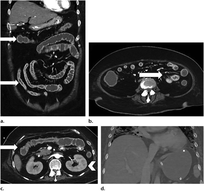
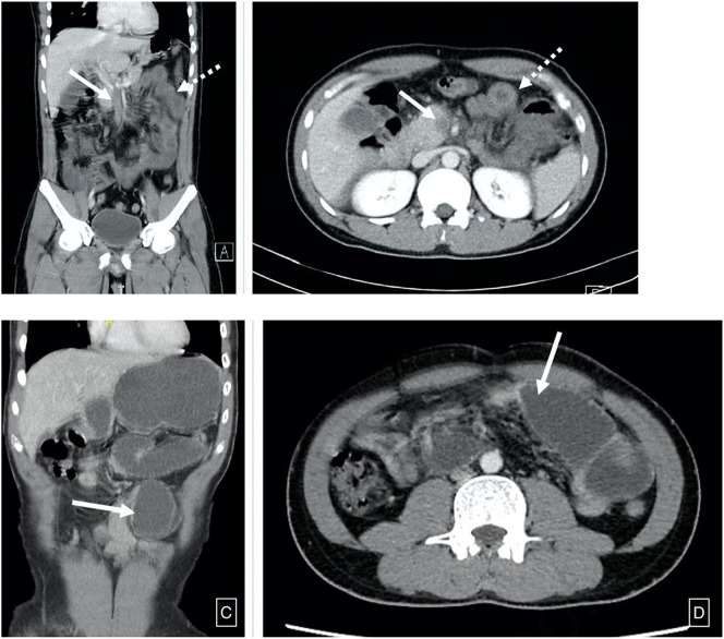
Background: Limited data is available addressing gastrointestinal (GI) ischemia in coronavirus disease 2019 (COVID-19). We reviewed the clinical and radiologic features of GI ischemia and its related complications in thirty-one COVID-19 patients reported in literature.

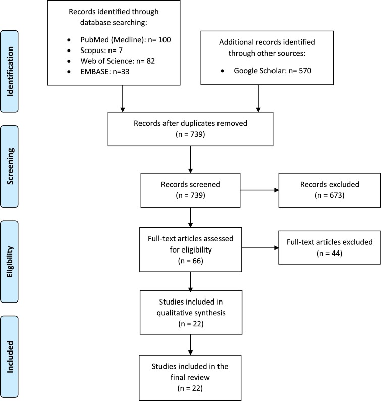
Methods: A systematic literature review was performed using a search strategy on all studies published from January 1, 2020, to June 13, 2020, and updated on September 6, 2020, on databases from PubMed, Scopus, Embase, Web of Science, and Google Scholar. Every study with at least one presentation of COVID-19-related GI ischemia complication and one GI imaging finding was included.

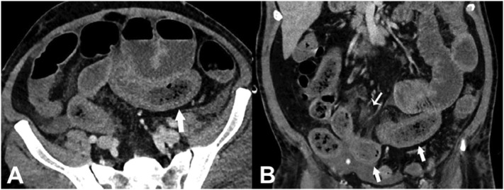
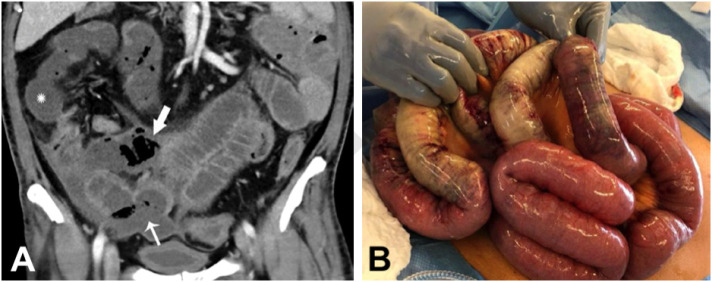
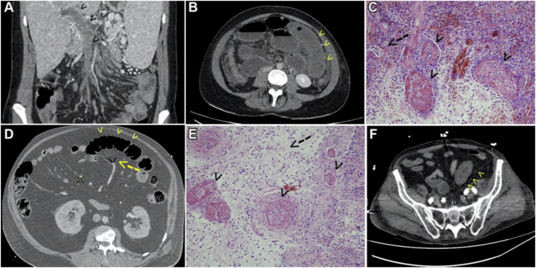
Results: In total, twenty-two studies and thirty-one patients with the mean age of 59 ± 12.7 (age range: 28-80) years old were included, of which 23 (74.2%) patients were male, 7 (22.5%) female, and one unknown gender. The significant GI imaging findings include mesenteric arterial or venous thromboembolism, followed by small bowel ischemia. Nine patients (29%) presented with arterial compromise due to superior mesenteric thromboembolism, resulting in bowel ischemia. Also, 6 patients (19.3%) demonstrated occlusive thrombosis of the portal system and superior mesenteric vein. More than two-thirds of patients (20, 64.5%) required laparotomy and bowel resection. Eventually, five (16.1%) patients were discharged, of whom four cases (12.9%) readmitted. Five (16.1%) patients remained ICU hospitalized at the report time and 12 (38.7%) patients died.

Conclusion: Macrovascular arterial/venous thrombosis is identified in almost half of COVID-19 patients with bowel ischemia. Overall mortality in COVID-19 patients with GI ischemia and radiologically evident mesenteric thrombotic occlusion was 38.7% and 40%, retrospectively.

Keywords: Abdominal pain; Coronavirus; Gastrointestinal; Ischemia; SARS-CoV-2; Tomography; X-ray, computed.


Conflict of interest statement

The authors of this manuscript declare no relationships with any companies, whose products or services may be related to the subject matter of the article.

Figures

Fig. 1
Fig. 1
Flow diagram of study selection process.
Fig. 2
Fig. 2
Axial (A) and coronal (B) CT of the abdomen and pelvis with IV contrast in a 57-year old man (patient no. 3) with a high clinical suspicion for bowel ischemia. There was generalized small bowel distension and segmental thickening (arrows), with adjacent mesenteric congestion (thin arrow in B), and a small volume of ascites (* in B). Findings are nonspecific but suggestive of early ischemia or infection. Images obtained from Bhayana et al. Radiology, published online May 11, 2020, copyright (2020), and permission to use granted by Ashley E. Daly, Senior Manager, Journal Rights & Communications Publications, Radiological Society of North America (RSNA).
Fig. 3
Fig. 3
Coronal (A) CT of the abdomen and pelvis with IV contrast in a 47-year-old man (patient no. 4) with abdominal tenderness demonstrates typical findings of mesenteric ischemia and infarction, including pneumatosis intestinalis (arrow) and non-enhancing bowel (*). Frank discontinuity of a thickened loop of small bowel in the pelvis (thin arrow) is in keeping with perforation. These findings were confirmed at laparotomy (B), with the additional observation of atypical yellow discoloration of bowel. Images obtained from Bhayana et al. Radiology, published online May 11, 2020, copyright (2020), and permission to use granted by Ashley E. Daly, Senior Manager, Journal Rights & Communications Publications, Radiological Society of North America (RSNA). (For interpretation of the references to color in this figure legend, the reader is referred to the web version of this article.)
Fig. 4
Fig. 4
Abdominal CT scan and pathological findings in SARS-CoV-2 infected patients. (A) CT scan shows mesenteric and portal vein thrombosis (arrowheads) in a young female patient (patient no. 12) with essential thrombocythemia, as a first sign and prior to respiratory symptoms revealing the COVID-19. (B) CT scan shows bowel infarction in the first bowel loop (arrowheads) 5 days after admission of the patient no. 12, prompting emergency laparotomy and bowel resection. (C) Pathological findings of patient no. 12 (hematoxylin and eosin stain, 200 magnification): small bowel necrosis; arrowheads show microthrombi in the lamina propria and the submucosa; arrow shows glandular necrosis. (D) CT scan shows signs of bowel ischemia (arrowheads) and mesenteric venous gas (arrow) in the proximal jejunum in a 56-year-old male patient (patient no. 13) with acute respiratory distress syndrome during COVID-19. (E) Pathological findings of patient no. 13 (hematoxylin and eosin stain, 200 magnification): small bowel necrosis; arrowheads show micro-thrombi; and arrow shows edema and inflammatory infiltrates in the submucosa. (F) CT scan shows an inflammatory bowel loop with thickening and edema (arrowhead) in a 67-year-old male (patient no. 14) with acute respiratory distress syndrome during COVID-19. Images obtained from Ignat. Surgery published on July 1, 2020, Vol. 168 (1) P14–16 (copyright 2020), and permission to use granted by Elsevier and Copyright Clearance Center.
Fig. 5
Fig. 5
Cough and shortness of breath in a 46-year-old woman (patient no. 22) with diabetes mellitus who was initially diagnosed with COVID-19. The patient's condition clinically deteriorated, and the use of mechanical ventilation, extracorporeal membrane oxygenation (ECMO), and pressor support was required. The clinical course was complicated by acute toxic metabolic encephalopathy, pneumonia, acute respiratory distress syndrome, cardiogenic shock, acute kidney injury, and secondary adrenal insufficiency. Coronal (a) and axial (b, c) contrast-enhanced CT images show multifocal small and large bowel wall thickening (arrows) creating a ribbonlike appearance, likely related to small vessel ischemia. Note the altered enhancement of the kidneys (arrowhead in c) in the setting of acute kidney injury and multifocal peripheral airspace opacities at the lung bases on the coronal CT image (d). Images obtained from Olson et al. Radiology published online on July 10, 2020, Vol. 40, No. 5, copyright (2020), and permission to use granted by Ashley E. Daly, Senior Manager, Journal Rights & Communications Publications, Radiological Society of North America (RSNA).
Fig. 6
Fig. 6
A computed tomography scan of the abdomen and pelvis revealed A, B: Superior mesenteric vein thrombosis (bold arrow) and diffuse mural thickening (dotted arrow), C, D: High grade small bowel obstruction with a transition point (bold arrow) seen in mid jejunum. Images obtained from Pang et al. Annals of Vascular Surgery available online on August 28, 2020, (copyright 2020), and permission to use granted by Elsevier and Copyright Clearance Center.

References

    1. Guan W.-j., Ni Z.-y., Hu Y., et al. Clinical characteristics of coronavirus disease 2019 in China. N Engl J Med. 2020;382(18):1708–1720. doi: 10.1056/NEJMoa2002032. - DOI - PMC - PubMed
    1. Wu Z., McGoogan J.M. Characteristics of and important lessons from the coronavirus disease 2019 (COVID-19) outbreak in China: summary of a report of 72 314 cases from the Chinese Center for Disease Control and Prevention. Jama. 2020;323(13):1239–1242. doi: 10.1001/jama.2020.2648. - DOI - PubMed
    1. World Health Organization Coronavirus disease (COVID-2019) situation reports. https://www.who.int/emergencies/diseases/novel-coronavirus-2019/situatio...
    1. Wang D., Hu B., Hu C., et al. Clinical characteristics of 138 hospitalized patients with 2019 novel coronavirus–infected pneumonia in Wuhan, China. Jama. 2020;323(11):1061–1069. doi: 10.1001/jama.2020.1585. - DOI - PMC - PubMed
    1. Li Q., Guan X., Wu P., et al. Early transmission dynamics in Wuhan, China, of novel coronavirus–infected pneumonia. N Engl J Med. 2020;382(13):1199–1207. doi: 10.1056/NEJMoa2001316. - DOI - PMC - PubMed

Publication types