

Pathogenesis and contemporary diagnoses for lateral hip pain: a scoping review
- PMID: 33341914
- PMCID: PMC8298339
- DOI: 10.1007/s00167-020-06354-1
Pathogenesis and contemporary diagnoses for lateral hip pain: a scoping review
Abstract
Purpose: Recent advances in diagnostic imaging techniques and soft tissue endoscopy now allow for precise diagnosis and management of extra-articular hip pathology. The aim of this scoping review is to present an evidence-based update of the relevant literature focussing only on the pathoanatomy, clinical assessment and the diagnosis of pathology in the peritrochanteric space.
Methods: A literature search was performed on PubMed to include articles which reported on the anatomy and diagnosis of greater trochanteric pain syndrome, trochanteric bursitis, gluteus medius tears and external snapping hip syndrome.
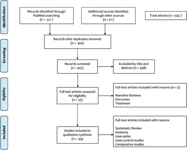
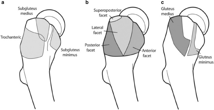
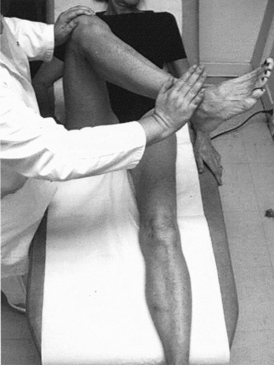
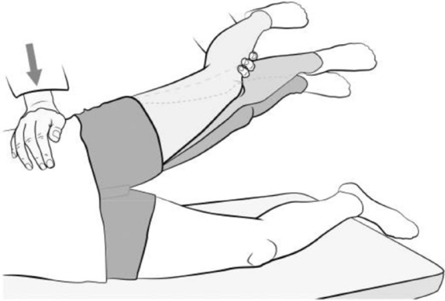
Results: A total of 542 studies were identified, of which 49 articles were included for full text analysis for the scoping review. Peritrochanteric space pathology can be broadly classified into (1) greater trochanteric pain syndrome (GTPS), (2) abductor tears and (3) external snapping hip syndrome. Anatomically, gluteus medius, gluteus minimus and tensor fascia lata work in conjunction to abduct and internally rotate the hip. The anterolateral part of the gluteus medius tendon is more prone to tears due to a thin tendinous portion. Increased acetabular anteversion has also been shown to be associated with gluteal and trochanteric bursitis. In terms of clinical examination, tests which were found to be most useful for assisting in the diagnoses of lateral hip pain were the single-leg stance, resisted external derotation of the hip, hip lag sign and the Trendelenburg's test. Dynamic ultrasound along with guided injections and MRI scan do assist in differentiating the pathology and confirming the diagnosis in patients presenting with lateral hip pain. Finally, the assessment of baseline psychological impairment is essential in this group of patients to ensure outcomes are optimised.
Conclusion: Lateral hip pain used to be a poorly defined entity, but advances in imaging and interest in sports medicine have led to a better understanding of the pathology, presentation and management of this cohort of patients. A thorough appreciation of the anatomy of the abductor musculature, specific clinical signs and imaging findings will lead to an appropriate diagnosis being made and management plan instituted.
Level of evidence: IV.
Keywords: Abductor tear; External snapping hip syndrome; Greater trochanteric pain syndrome; Hip.
© 2020. The Author(s).
Conflict of interest statement
None related to this article. VK is an educational consultant for Smith & Nephew and Arthrex. AS has received payment for teaching activities from Stryker.
Figures

References
-
- Allen WC, Cope R. Coxa saltans: the snapping hip revisited. J Am Acad Orthop Surg. 1995;3(5):303–308. - PubMed
-
- Audenaert E, Pattyn C. Balloon dissection for improved access to the peritrochanteric compartment. Arthroscopy. 2009;25(11):1349–1353. - PubMed
-
- Beck M, Sledge JB, Gautier E, Dora CF, Ganz R. The anatomy and function of the gluteus minimus muscle. J Bone Joint Surg Br. 2000;82(3):358–363. - PubMed
-
- Beighton P, Horan F. Orthopaedic aspects of the Ehlers-Danlos syndrome. J Bone Joint Surg Br. 1969;51(3):444–453. - PubMed
-
- Blankenbaker DG, Ullrick SR, Davis KW, De Smet AA, Haaland B, Fine JP. Correlation of MRI findings with clinical findings of trochanteric pain syndrome. Skeletal Radiol. 2008;37(10):903–909. - PubMed
Publication types
MeSH terms
LinkOut - more resources
Full Text Sources
Research Materials
