. 2021 Oct;92(10):1483-1495.
doi: 10.1002/JPER.20-0529. Epub 2021 Jan 6.

Senescent cells exacerbate chronic inflammation and contribute to periodontal disease progression in old mice

Affiliations

Senescent cells exacerbate chronic inflammation and contribute to periodontal disease progression in old mice

Ruben Aquino-Martinez et al. J Periodontol. 2021 Oct.

Abstract

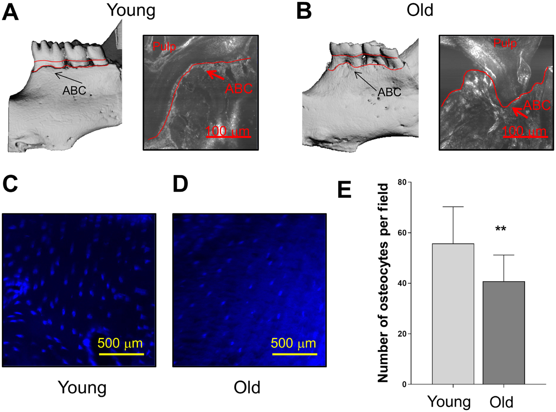
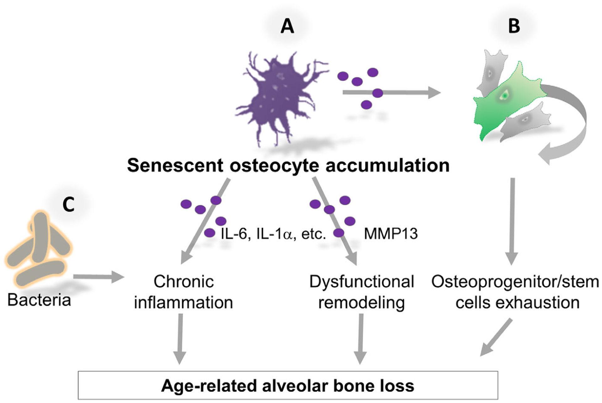
Background: Coinciding with other chronic comorbidities, the prevalence of periodontal disease increases with aging. Mounting evidence has established that senescent cells accumulate at sites of age-related pathologies, where they promote "non-microbial" inflammation. We hypothesized that alveolar bone osteocytes develop senescence characteristics in old age.

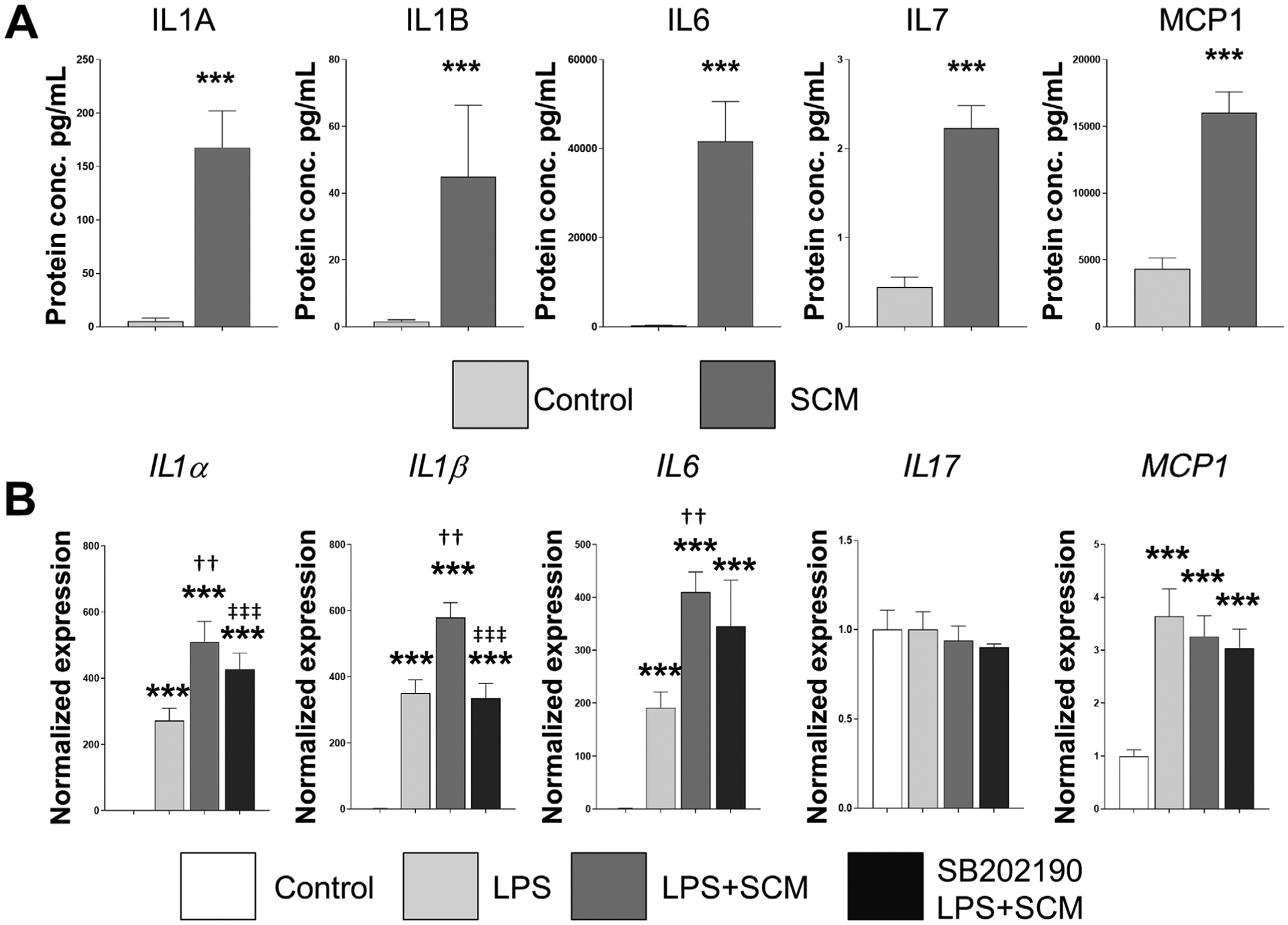
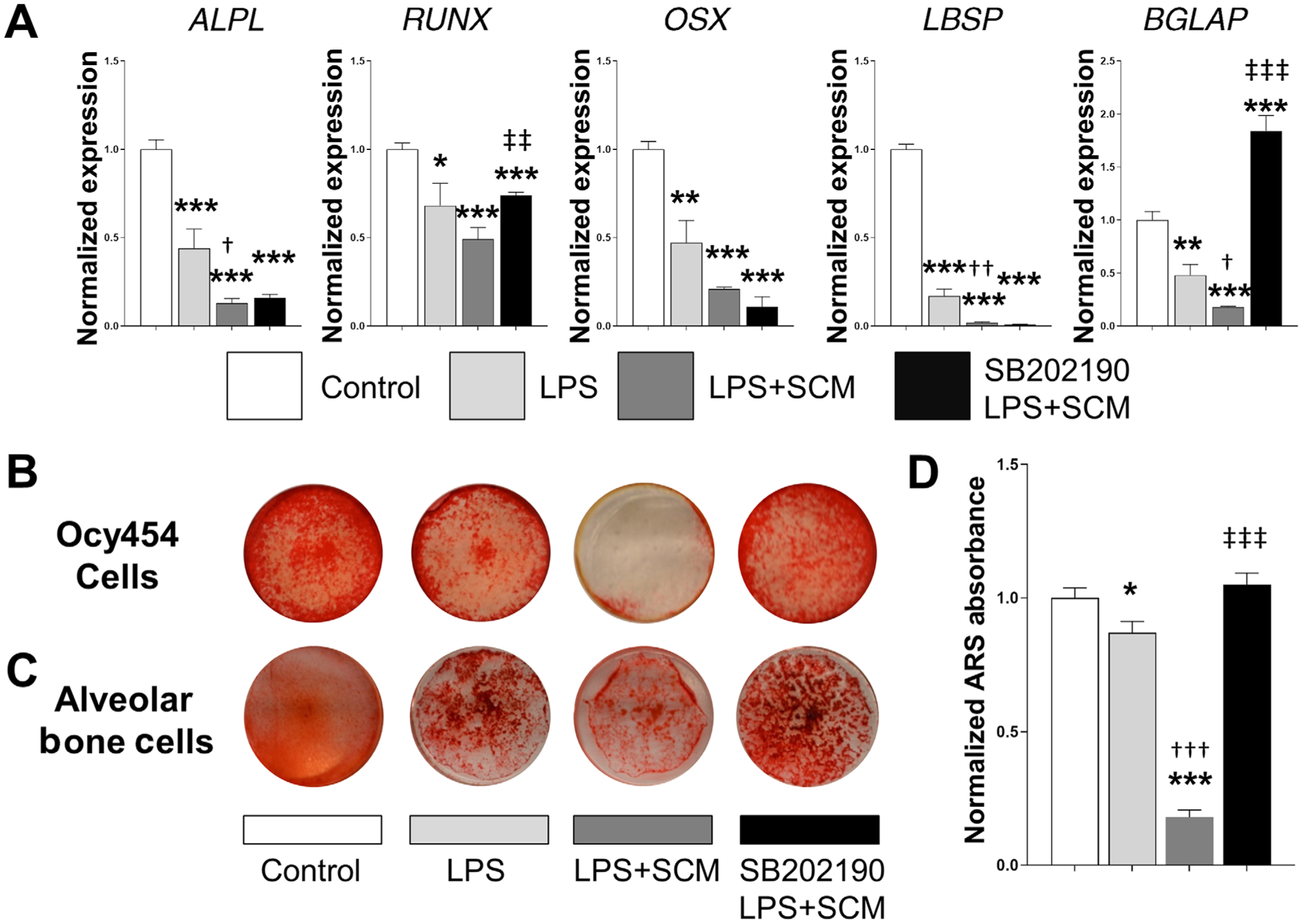
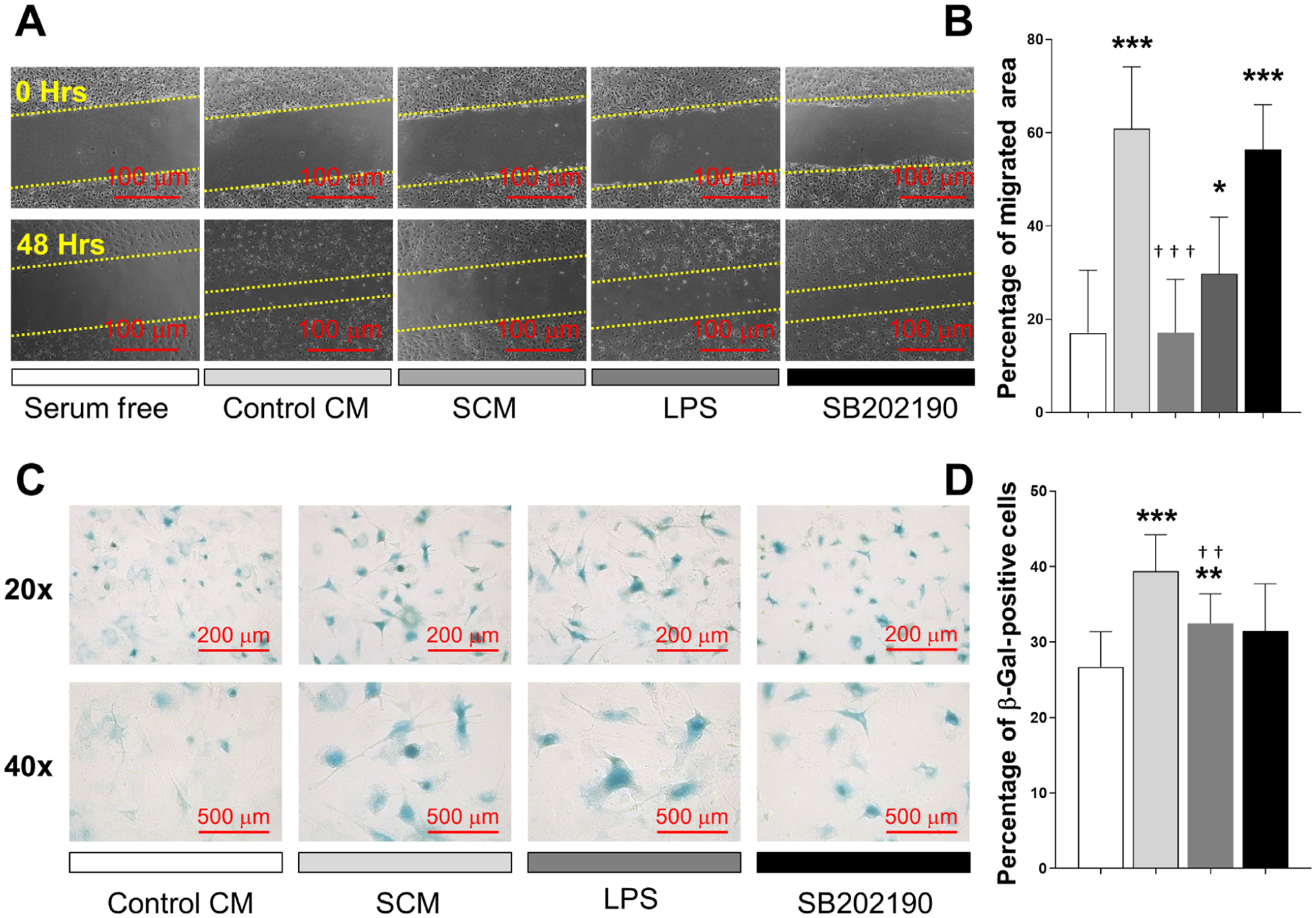
Methods: Alveolar bone samples were obtained from young (6 months) and old (20 to 22 months) mice to evaluate the expression of senescence biomarkers by immunofluorescent staining. Osteocyte-enriched fractions were used to characterize the age-related senescence-associated secretory phenotype (SASP) gene expression profile. Primary alveolar bone cells were exposed to the SASP via in vitro senescent conditioned media (SCM) administration. A multiplex assay confirmed protein levels of specific cytokines. Interactions with bacterial components were evaluated by stimulating cells with lipopolysaccharide (LPS).

Results: Increased senescence-associated distension of satellites (SADS) and p16Ink4a mRNA expression were identified in alveolar bone osteocytes with aging. These findings were associated with increased levels of DNA damage, and activated p38 MAPK, both inducers of senescence. Furthermore, interleukin-6 (IL6), IL17, IGFBP4, and MMP13 were significantly upregulated with aging in osteocyte-enriched samples. Interestingly, SCM potentiated the LPS-induced expression of IL1α, IL1β, and IL6. Cell migration and differentiation were also impeded by SCM. These in vitro effects were ameliorated by the p38 MAPK inhibitor SB202190.

Conclusions: Accumulation of senescent osteocytes contributes to deterioration of the periodontal environment by exacerbating chronic inflammation and reducing regeneration in old age. Cellular senescence is a cell-intrinsic response to DNA damage, and a host-related mechanism associated with aging that could potentiate inflammation induced by bacterial components.

Keywords: DNA damage; aging; alveolar bone loss; cellular senescence; inflammation; periodontal diseases.


Figures

FIGURE 1
FIGURE 1
Osteocytes displaying senescent features accumulate in alveolar bone with aging. Expression of established senescence biomarkers were evaluated in alveolar bone samples from 6-month-old and 20- to 22-month-old wild-type mice. A and B) Representative images of normal and senescent alveolar bone osteocytes using the SADS assay (see white arrows for distended DNA satellites); magnification 100×. C) Percentage of alveolar bone osteocytes displaying SADS (from Panels A-B). D) γH2AX immunofluorescence signal (red) showing age-dependent DNA damage. E) Images displaying p53 signal (green). F) The DNA damage pathway p38 MAPK is displayed in green. Nuclei were stained with DAPI. G) p16Ink4a, p21, and p53 expression from osteocyte-enriched populations isolated from young and old alveolar bone without in vitro cell culture (n = 10). H) Osteocyte-enriched fraction from old alveolar bone exhibited upregulated expression of the following factors: IGFBP4, IL6, IL17, and MMP13. Data represent mean ± SEM. Significant changes are indicated by *P < 0.05; **P < 0.01; ***P < 0.001 relative to young versus old samples
FIGURE 2
FIGURE 2
Senescence-associated factors interact with LPS. A) Jawbone cells isolated from 6-month-old mice were differentiated into osteocytes, by repeatedly exposed to LPS (10 μg/mL). CM from these senescent and healthy osteocytes was collected and analyzed by a bead-based multiplex cytokine array. Six replicates were used for each condition and protein concentration displayed in pg/mL. B) To evaluate the potential interaction between senescence-associated factors and LPS, differentiated osteocytes were exposed to media plus control CM (control), LPS (10 μg/mL) or LPS combined with SCM for 48 hours. The effect of SB202190, a selective p38 MAPK inhibitor (10 μM), was also evaluated by pretreating cells before starting the experiments. Data represent mean ± SEM. Significant changes are indicated by: * compared with control, † LPS compared with LPS+SCM, and ‡ LPS+SCM compared with LPS+SCM+SB202190. Various levels of significance are based on the number of each respective symbol (one symbol P < 0.05; two symbols P < 0.01; three symbols P < 0.001)
FIGURE 3
FIGURE 3
Age-related alveolar bone loss is associated with decreased osteocyte number. Representative μCT and bright-field confocal images display age-related alveolar bone loss comparing (A) 6-month-old and (B) 20- to 22-month-old mice. Increased distance from the cemento-enamel junction to alveolar bone crest (ABC) is observed in the old mice (red lines). C and D) Representative alveolar bone sections from 6- and 20- to 22-month-old wild-type mice were stained with DAPI. E) The results are presented as average number of osteocytes per field. Significant changes are indicated by **P < 0.01
FIGURE 4
FIGURE 4
Senescent osteocyte-secreted factors aggravate osteocyte differentiation and mineralization induced by LPS. A) Differentiated Ocy454 cells were exposed to the various stimuli shown in the figure and described in the Methods, and the expression of recognized osteogenic markers was measured by RT-qPCR. Data are represented as mean ± SEM (n = 6). B) A subset of cells from panel A was used to assess bone mineralization capacity using ARS staining. C) A parallel experiment was performed using primary alveolar osteocytic cells. They were cultured under the same conditions for 15 days, and mineralized nodule formation evaluated using ARS staining. Note that in both experiments decreased mineralization produced by LPS is notably aggravated by the addition of SCM, which is reversed by SB202190. Representative images for both cell models were obtained using a digital camera. D) ARS elution was performed on the Ocy454 cells (n = 6) and normalized ARS absorbance is displayed. Significant changes are indicated by: * compared with control, †LPS compared with LPS+SCM, and ‡ for LPS+SCM compared with LPS+SCM+SB202190. Various levels of significance are based on the number of each respective symbol (one symbol P < 0.05; two symbols P < 0.01; three symbols P < 0.001)
FIGURE 5
FIGURE 5
Senescent osteocyte secreted factors induce a deleterious effect on osteoprogenitor cells. A and B) A wound healing assay was used to measure the effect of SCM on chemotactic cell recruitment. Cells were exposed to serum free media as control, CM from healthy osteocytes (control CM), SCM from irradiated osteocytes, LPS (10 μg/mL), or SCM plus SB202190 (10 μM). Cells were allowed to migrate into the scratched area for 48 hours. Representative images from each condition are displayed, and results presented as percentage of migrated area. C and D) The acquisition of senescence-associated SA-β-activity was evaluated by treating primary cells with control CM, SCM, LPS (10 μg/mL) or SCM combined with SB202190. Representative images from each condition are displayed and the results are presented as percentage of β-gal positive cells. Significant changes are indicated by: * compared with serum free or control CM, and † compared with control CM versus SCM or SCM versus SB202190. Various levels of significance are based on the number of each respective symbol (one symbol P < 0.05; two symbols P < 0.01; three symbols P < 0.001)
FIGURE 6
FIGURE 6
Hypothetical mechanism by which senescent cells deteriorate the old periodontal environment. A) Senescent osteocytes accumulate in alveolar bone with aging as a DNA damage response and as result of impaired clearance by the immune system. Senescent osteocytes-associated proinflammatory and proteolytic factors (purple circles) promote exacerbated local inflammation and dysfunctional bone extracellular matrix remodeling. B) The impact of both paracrine and age-related replicative senescence on stem cells contributes to the exhaustion of osteoprogenitor and stem cells. Thus, accumulation of senescent osteocytes deteriorates the periodontal environment and contributes to alveolar bone destruction with age. C) Senescent cells are an additional source of proinflammatory cytokines that could exacerbate bacterial-induced inflammation

References

    1. Franceschi C, Campisi J. Chronic inflammation (inflammaging) and its potential contribution to age-associated diseases. J Gerontol A Biol Sci Med Sci. 2014;69Suppl 1:S4–S9. - PubMed
    1. Lopez-Otin C, Blasco MA, Partridge L, Serrano M, Kroemer G. The hallmarks of aging. Cell. 2013;153:1194–1217. - PMC - PubMed
    1. Kamer AR, Pirraglia E, Tsui W, et al.Periodontal disease associates with higher brain amyloid load in normal elderly. Neurobiol Aging. 2015;36:627–633. - PMC - PubMed
    1. Wang CJ, McCauley LK. Osteoporosis and Periodontitis. Curr Osteoporos Rep. 2016;14:284–291. - PMC - PubMed
    1. Ebersole JL, Graves CL, Gonzalez OA, et al.Aging, inflammation, immunity and periodontal disease. Periodontol 2000. 2016;72:54–75. - PubMed

Publication types