. 2020 Jan-Dec:16:1744806920970099.
doi: 10.1177/1744806920970099.

IPSE, a parasite-derived, host immunomodulatory infiltrin protein, alleviates resiniferatoxin-induced bladder pain

Affiliations

IPSE, a parasite-derived, host immunomodulatory infiltrin protein, alleviates resiniferatoxin-induced bladder pain

Kenji Ishida et al. Mol Pain. 2020 Jan-Dec.

Abstract

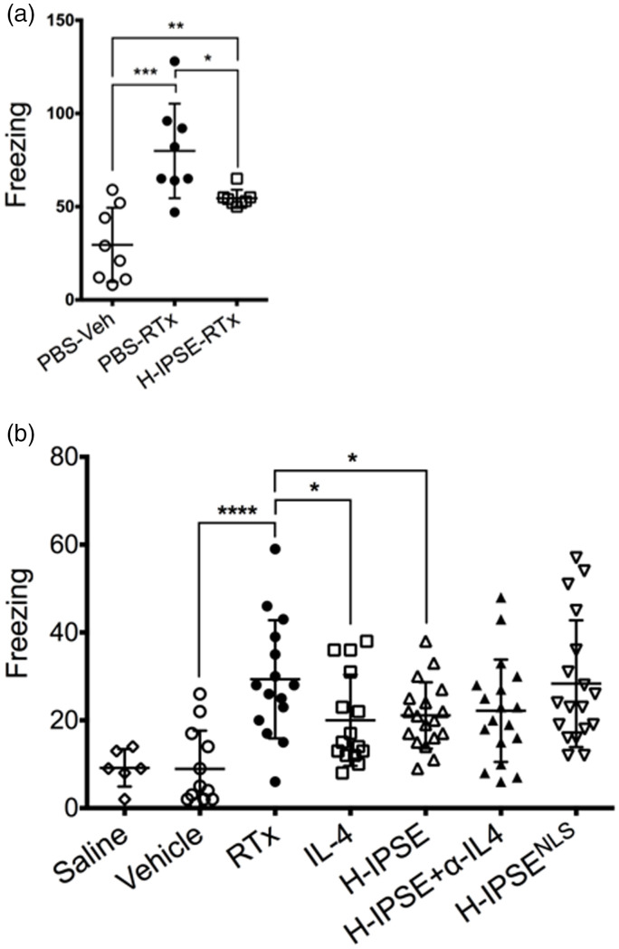
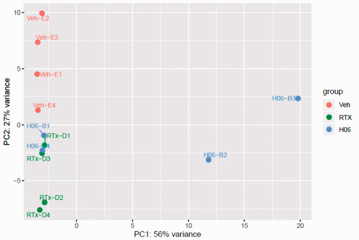
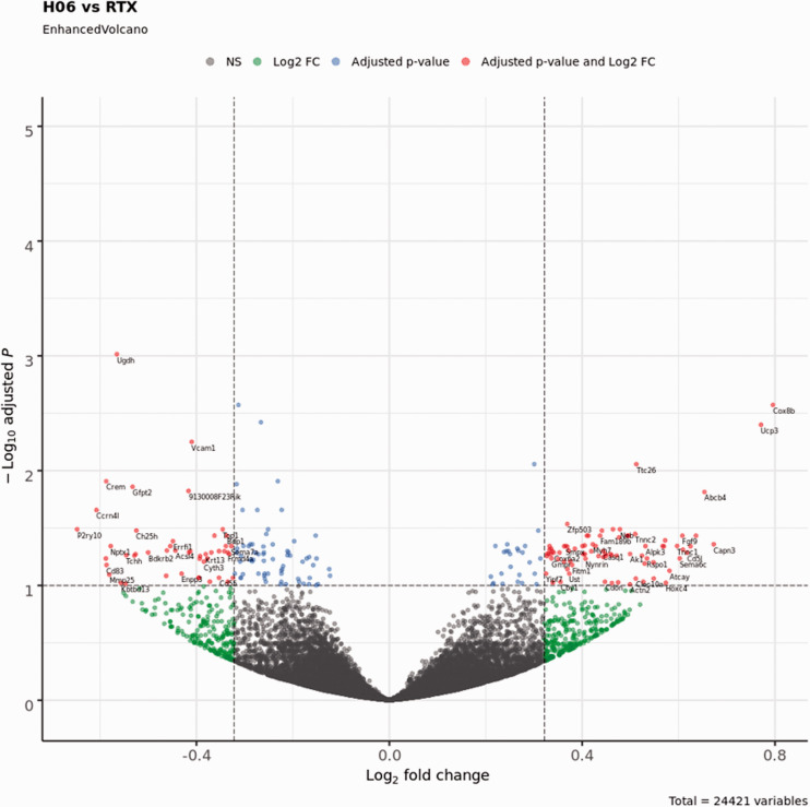
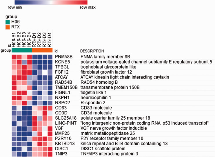
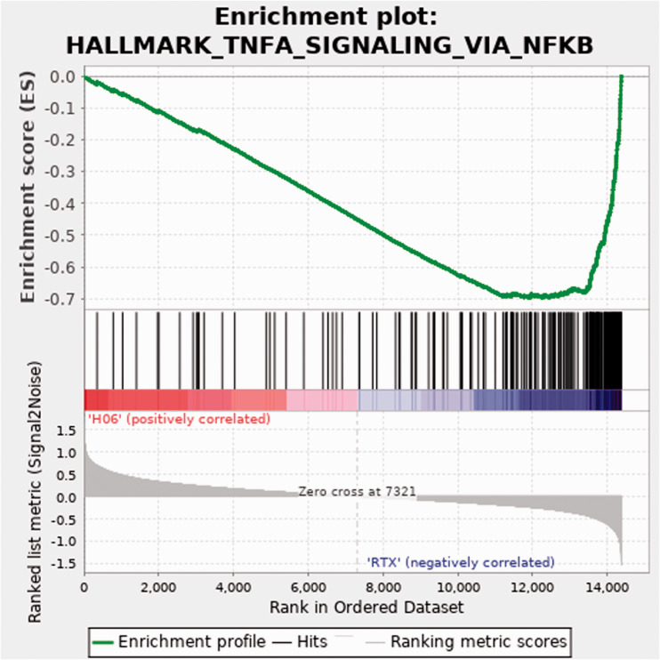
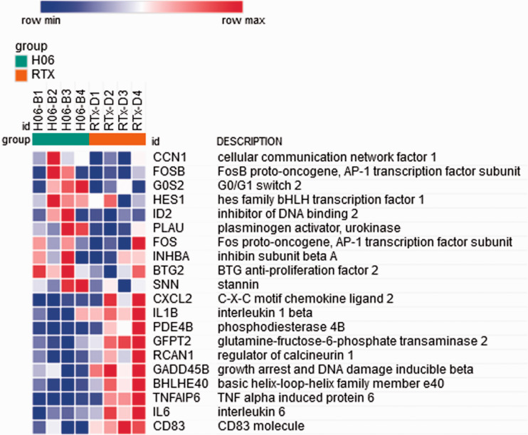
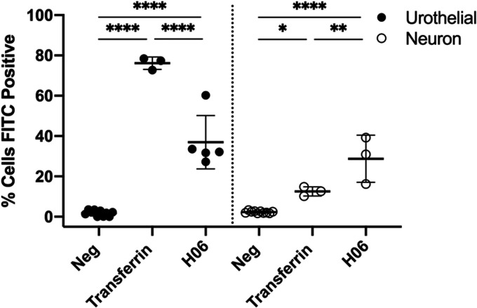
The transient receptor potential cation channel subfamily V member 1 (TRPV1) receptor is an important mediator of nociception and its expression is enriched in nociceptive neurons. TRPV1 signaling has been implicated in bladder pain and is a potential analgesic target. Resiniferatoxin is the most potent known agonist of TRPV1. Acute exposure of the rat bladder to resiniferatoxin has been demonstrated to result in pain-related freezing and licking behaviors that are alleviated by virally encoded IL-4. The interleukin-4-inducing principle of Schistosoma mansoni eggs (IPSE) is a powerful inducer of IL-4 secretion, and is also known to alter host cell transcription through a nuclear localization sequence-based mechanism. We previously reported that IPSE ameliorates ifosfamide-induced bladder pain in an IL-4- and nuclear localization sequence-dependent manner. We hypothesized that pre-administration of IPSE to resiniferatoxin-challenged mice would dampen pain-related behaviors. IPSE indeed lessened resiniferatoxin-triggered freezing behaviors in mice. This was a nuclear localization sequence-dependent phenomenon, since administration of a nuclear localization sequence mutant version of IPSE abrogated IPSE's analgesic effect. In contrast, IPSE's analgesic effect did not seem IL-4-dependent, since use of anti-IL-4 antibody in mice given both IPSE and resiniferatoxin did not significantly affect freezing behaviors. RNA-Seq analysis of resiniferatoxin- and IPSE-exposed bladders revealed differential expression of TNF/NF-κb-related signaling pathway genes. In vitro testing of IPSE uptake by urothelial cells and TRPV1-expressing neuronal cells showed uptake by both cell types. Thus, IPSE's nuclear localization sequence-dependent therapeutic effects on TRPV1-mediated bladder pain may act on TRPV1-expressing neurons and/or may rely upon urothelial mechanisms.

Keywords: Bladder; analgesic; immune modulation; pain; parasite; schistosoma.


Conflict of interest statement

Declaration of Conflicting Interests: The author(s) declared no potential conflicts of interest with respect to the research, authorship, and/or publication of this article.

Figures

Figure 1.
Figure 1.
IPSE reduces resiniferatoxin-induced, pain-related freezing behaviors in an IL-4- and nuclear localization sequence-dependent manner. (a) Mice were administered intravenous phosphate-buffered saline (PBS) followed by intravesical PBS/Tween/ethanol vehicle (“PBS-Veh”), intravenous PBS with intravesical resiniferatoxin in vehicle (“PBS-RTx”), or one intravenous dose of the H06 H-IPSE ortholog of IPSE 24 hours before intravesical resiniferatoxin in vehicle (“H-IPSE-RTx”). (b) Mice were given intravenous PBS and intravesical PBS (“saline”), intravenous PBS and intravesical PBS/Tween/ethanol vehicle (“Vehicle”), intravenous PBS and intravesical resiniferatoxin in vehicle (“RTx”), recombinant IL-4 given intraperitoneally followed by intravesical resiniferatoxin in vehicle (“IL-4”), the H06 H-IPSE ortholog of IPSE given intravenously 24 hours before intravesical PBS with resiniferatoxin in vehicle (“H-IPSE”), the H06 H-IPSE ortholog of IPSE 24 hours given intravenously and anti-IL-4 antibody given intraperitoneally 30 minutes before resiniferatoxin in vehicle administered intravesically (“H-IPSE+α-IL4”), or a nuclear localization sequence (NLS) mutant of H06 H-IPSE given intravenously 24 hours before resiniferatoxin in vehicle administered intravesically (“H-IPSENLS”).
Figure 2.
Figure 2.
Principal component analysis of resiniferatoxin- and IPSE-treated bladder gene expression. Principal component analysis showed homogeneous clustering of gene expression among resiniferatoxin-treated mice (green symbols labeled with “RTx-D”) and vehicle-treated mice (red symbols labeled with “Veh-E”). There was some overlap of gene expression among resiniferatoxin-treated mice and mice treated with both H06 H-IPSE and resiniferatoxin (blue symbols labeled with “H06-B”).
Figure 3.
Figure 3.
Volcano plot showing differentially expressed genes between bladders treated with IPSE and resiniferatoxin (H06) versus bladders treated with resiniferatoxin alone (RTX). The cutoff value for the adjusted p-value was set at < 0.1, and the cutoff for the absolute value of the Log2 fold change was set at > 0.322 (1.25-fold in either direction). Blue dots represent genes satisfying the adjusted p-value cutoff. Green dots represent genes satisfying the Log2 fold change cutoff. Red dots represent genes satisfying both of these cutoff conditions and are labeled with the corresponding gene symbols. Gray dots represent genes that do not satisfy either condition (NS, not significant).
Figure 4.
Figure 4.
Heat map of genes from the 50 hallmark gene sets from the Molecular Signatures Database with enriched differential expression in bladders exposed to IPSE combined with resiniferatoxin or resiniferatoxin alone. The 10 most strongly differentially expressed genes associated with each treatment are shown. Each column shows gene expression for an individual mouse bladder. Green and red column coloring indicates IPSE combined with resiniferatoxin (H06) versus resiniferatoxin only (RTX)-treated bladders, respectively. Genes are sorted by signal-to-noise scores, and their symbols and names are listed in rows. Darkest blue to darkest red coloring represents lowest to highest gene expression, respectively, based on normalized read counts.
Figure 5.
Figure 5.
Enrichment plot for the TNF signaling via NF-κB pathway. Top panel, green line indicates running enrichment score for the TNF signaling via NF-κB pathway as the gene set enrichment analysis walks down the ranked list of genes. Middle panel depicts where the members of the TNF signaling via NF-κB pathway gene set appear in the ranked list of genes. Bottom panel shows the value of the ranking metric moving down the list of ranked genes. Positive values indicate correlation with the first phenotype (H06; H-IPSE combined with resiniferatoxin) and negative values indicate correlation with the second phenotype (RTX; resiniferatoxin alone).
Figure 6.
Figure 6.
Heat map of gene members of the TNF signaling via NF-κB pathway with enriched differential expression in bladders exposed to IPSE combined with resiniferatoxin or resiniferatoxin alone. The 10 most strongly differentially expressed genes associated with each treatment are shown. Each column shows gene expression for an individual mouse bladder. Green and red column coloring indicates IPSE combined with resiniferatoxin (H06) versus resiniferatoxin only (RTX)-treated bladders, respectively. Genes are sorted by signal-to-noise scores, and their symbols and names are listed in rows. Darkest blue to darkest red coloring represents lowest to highest gene expression, respectively, based on normalized read counts.
Figure 7.
Figure 7.
Internalization of IPSE by neuronal and urothelial cells. Cath.a mouse neuronal and HCV-29 human urothelial cells were incubated for 16 hrs with Alexa 488 conjugated H06 H-IPSE (1 μg/ml) or transferrin (4 μg/ml) and analyzed by flow cytometry after trypan blue quenching of extracellular Alexa 488 signal. Data is representative of 2 experiments. *p = 0.0139, **p = 0.0025, ****p<0.0001.

Similar articles

Cited by

References

    1. Elliott TR. The innervation of the bladder and urethra. J Physiol 1907; 35: 367–445. - PMC - PubMed
    1. De Groat WC, Yoshimura N. Afferent nerve regulation of bladder function in health and disease. Handb Exp Pharmacol 2009; 194: 91–138. - PMC - PubMed
    1. Schramm G, Gronow A, Knobloch J, Wippersteg V, Grevelding CG, Galle J, Fuller H, Stanley RG, Chiodini PL, Haas H, Doenhoff MJ. IPSE/alpha-1: a major immunogenic component secreted from Schistosoma mansoni eggs. Mol Biochem Parasitol 2006; 147: 9–19. - PubMed
    1. Kaur I, Schramm G, Everts B, Scholzen T, Kindle KB, Beetz C, Montiel-Duarte C, Blindow S, Jones AT, Haas H, Stolnik S, Heery DM, Falcone FH. Interleukin-4-inducing principle from Schistosoma mansoni eggs contains a functional C-terminal nuclear localization signal necessary for nuclear translocation in mammalian cells but not for its uptake. Infect Immun 2011; 79: 1779–1788. - PMC - PubMed
    1. Mbanefo EC, Le L, Zee R, Banskota N, Ishida K, Pennington LF, Odegaard JI, Jardetzky TS, Alouffi A, Falcone FH, Hsieh MH. IPSE, a urogenital parasite-derived immunomodulatory protein, ameliorates ifosfamide-induced hemorrhagic cystitis through downregulation of pro-inflammatory pathways. Sci Rep 2019; 9: 1–13. - PMC - PubMed

Publication types

MeSH terms