Meta-Analysis
. 2021 Feb:115:107682.
doi: 10.1016/j.yebeh.2020.107682. Epub 2020 Dec 4.

Meta-analysis of EEG findings in patients with COVID-19

Affiliations
Meta-Analysis

Meta-analysis of EEG findings in patients with COVID-19

Takafumi Kubota et al. Epilepsy Behav. 2021 Feb.

Abstract

Objective: To perform a systematic review and meta-analysis to summarize and quantitatively evaluate the electroencephalogram (EEG) findings in patients with coronavirus disease 2019 (COVID-19).

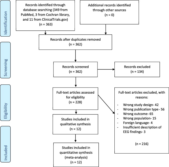
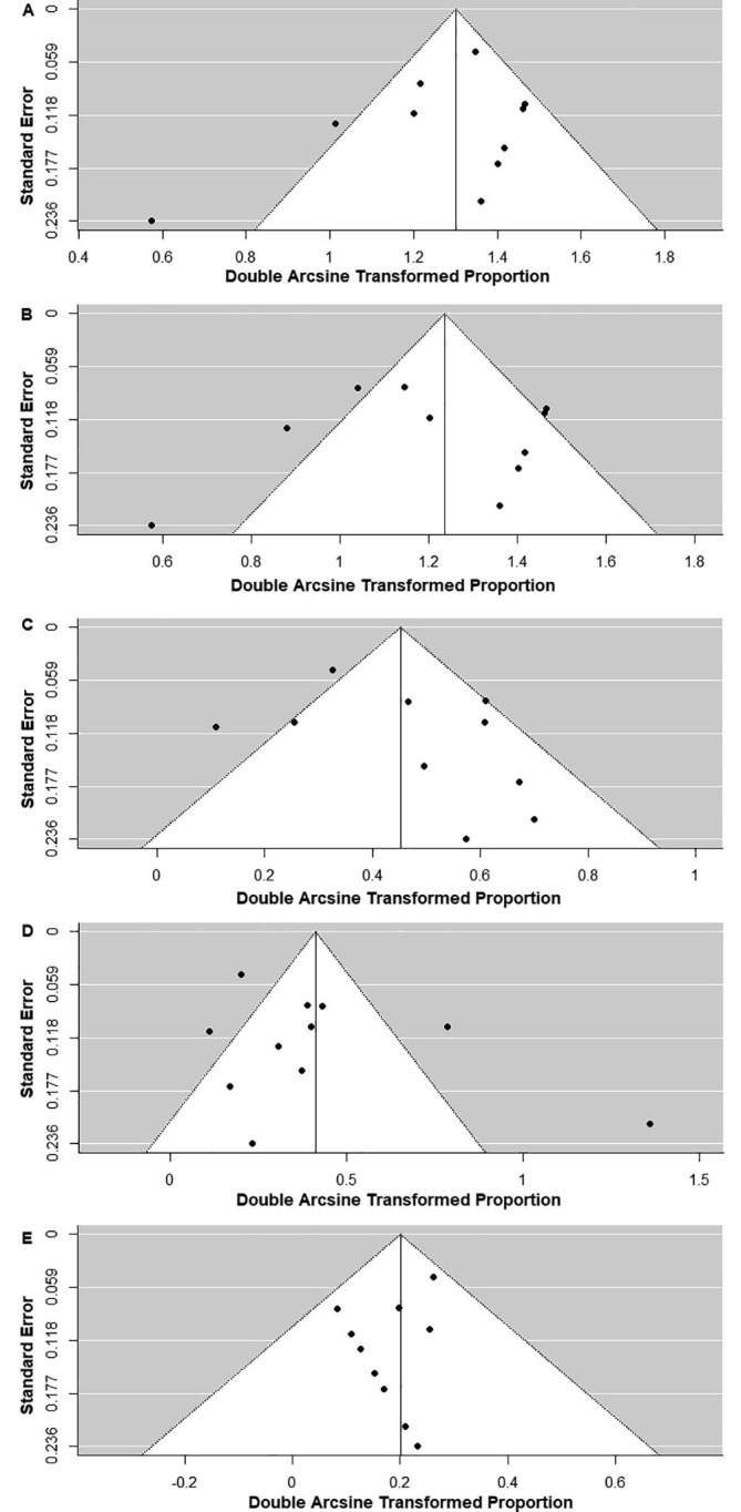
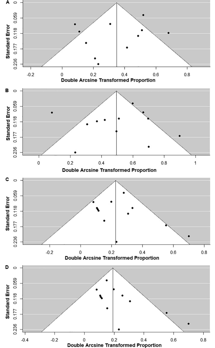
Methods: The MEDLINE, CENTRAL, and ClinicalTrials.Gov databases were comprehensively assessed and searched for observational studies with EEG findings in patients with COVID-19. Pooled proportions of EEG findings with 95% confidence intervals (CIs) were assessed using a random effects model. The quality of assessment for each study, heterogeneity between the studies, and publication bias were also evaluated.

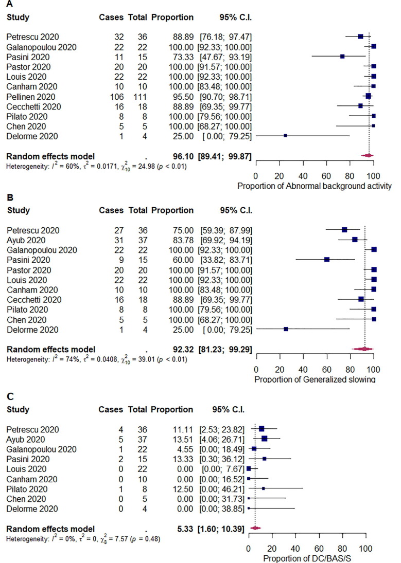
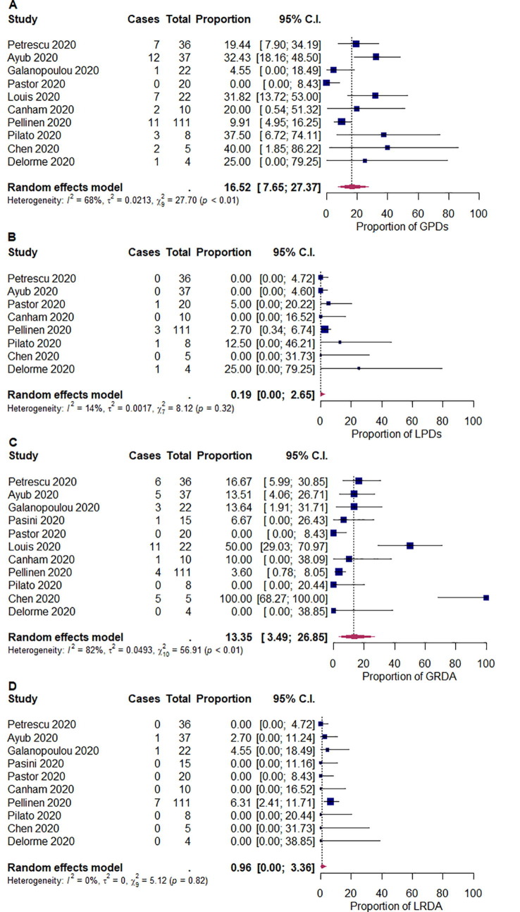
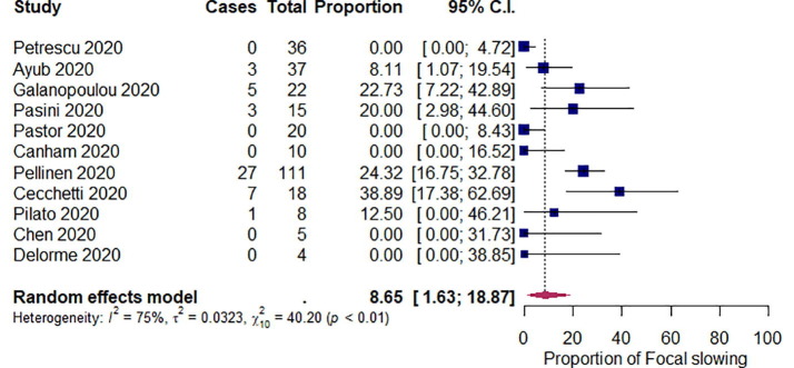
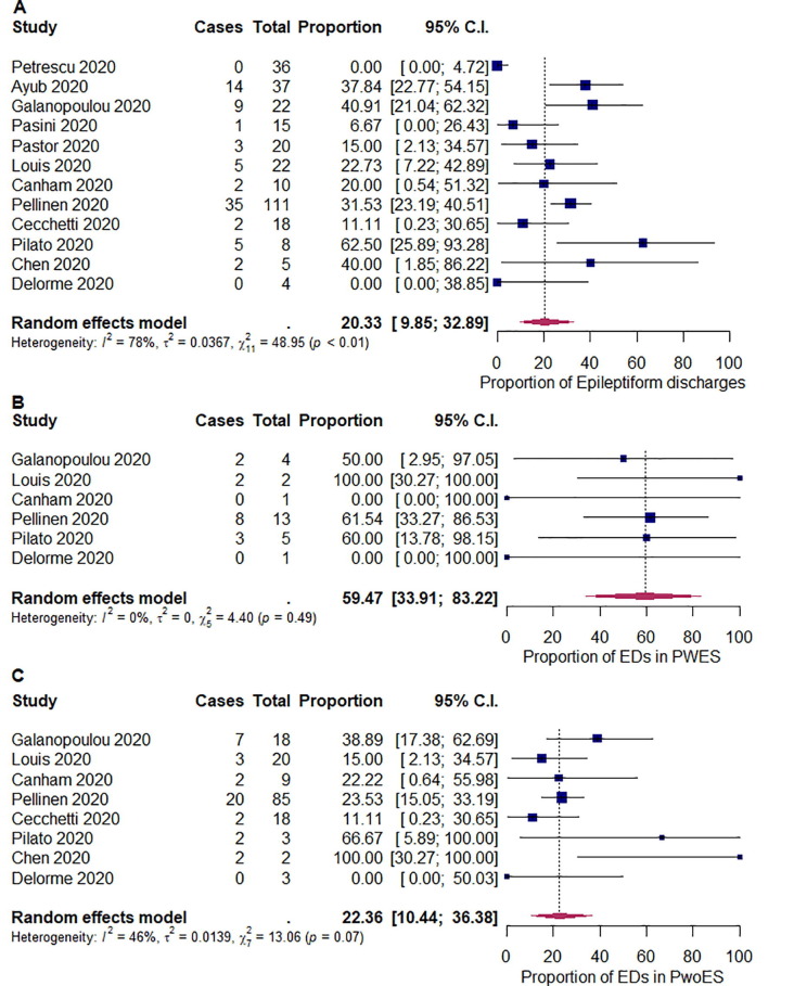
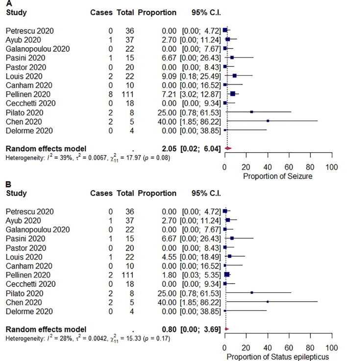
Results: In total, 12 studies with 308 patients were included in the meta-analysis. Abnormal background activity and generalized slowing in the pooled proportions were common findings among the patients with COVID-19 (96.1% [95% CI: 89.4-99.9]; I2 = 60%; p < 0.01 and 92.3% [95% CI: 81.2-99.3]; I2 = 74%; p < 0.01, respectively). The proportion of patients with epileptiform discharges (EDs) was 20.3% ([95% CI: 9.85-32.9]; I2 = 78%; p < 0.01). The proportion of EDs varied between patients with a history of epilepsy or seizures (59.5% [95% CI: 33.9-83.2]; I2 = 0%; p = 0.49) and patients without them (22.4% [95% CI: 10.4-36.4]; I2 = 46%; p = 0.07). The findings of seizures and status epilepticus on EEG were observed in 2.05% ([95% CI: 0.02-6.04]; I2 = 39%; p = 0.08) and 0.80% ([95% CI: 0.00.-3.69]; I2 = 28%; p = 0.17) of the patients, respectively.

Conclusion: The proportion of abnormal background activity in patients with COVID-19 was high (96.1%). Epileptiform discharges were present in 20.3% of the cases and the proportion varied between people who had a history of epilepsy/seizure and those who did not. However, the proportion of seizures and status epilepticus on EEG was low (2.05% and 0. 80%, respectively).

Keywords: COVID-19; EEG; Epilepsy; Neurology; Seizure; Status epilepticus.


Figures

Fig. 1
Fig. 1
Preferred reporting items for systematic reviews and meta-analyses (PRISMA) flow diagram of this research.
Fig. 2
Fig. 2
Random-effects meta-analysis of the pooled proportions of (A) abnormal background activity, (B) generalized slowing, and (C) discontinuous/burst-attenuation or suppression/suppression (DC/BAS/S). 95% C.I. = 95% confidence intervals.
Fig. 3
Fig. 3
Random-effects meta-analysis of the pooled proportions of (A) generalized periodic discharges (GPDs), (B) lateralized periodic discharges (LPDs), (C) generalized rhythmic delta activity (GRDA), and (D) lateralized rhythmic delta activity (LRDA). 95% C.I. = 95% confidence intervals.
Fig. 4
Fig. 4
Random-effects meta-analysis of the pooled proportion of focal slowing. 95% C.I. = 95% confidence intervals.
Fig. 5
Fig. 5
Random-effects meta-analysis of the pooled proportions of (A) epileptiform discharges (EDs), (B) EDs in patients with a history of epilepsy or seizure (PWES), and (C) EDs in patients without a history of epilepsy or seizure (PwoES). 95% C.I. = 95% confidence intervals.
Fig. 6
Fig. 6
Random-effects meta-analysis of the pooled proportions of (A) electrographic seizure and (B) electrographic status epilepticus. 95% C.I. = 95% confidence intervals.
Fig. 7
Fig. 7
Funnel plots of the meta-analysis of (A) abnormal background activity, (B) generalized slowing, (C) generalized periodic discharges, (D) generalized rhythmic delta activity, and (E) lateralized rhythmic delta activity.
Fig. 8
Fig. 8
Funnel plots of the meta-analysis of (A) focal slowing, (B) epileptiform discharges, (C) electrographic seizure, and (D) electrographic status epilepticus.

References

    1. Guan W., Ni Z., Hu Y.H.Y.Y.H., Liang W., Ou C., He J., et al. Clinical characteristics of coronavirus disease 2019 in China. N Engl J Med. 2020;382:1708–1720. doi: 10.1056/NEJMoa2002032. - DOI - PMC - PubMed
    1. Mao L., Jin H., Wang M., Hu Y., Chen S., He Q., et al. Neurologic manifestations of hospitalized patients with coronavirus disease 2019 in Wuhan, China. JAMA Neurol. 2020 doi: 10.1001/jamaneurol.2020.1127. - DOI - PMC - PubMed
    1. Varatharaj A., Thomas N., Ellul M.A., Davies N.W.S., Pollak T.A., Tenorio E.L., et al. Neurological and neuropsychiatric complications of COVID-19 in 153 patients: a UK-wide surveillance study. Lancet Psychiatry. 2020 - PMC - PubMed
    1. Ahmed M.U., Hanif M., Ali M.J., Haider M.A., Kherani D., Memon G.M., et al. Neurological manifestations of COVID-19 (SARS-CoV-2): a review. Front Neurol. 2020;11:518. doi: 10.3389/fneur.2020.00518. - DOI - PMC - PubMed
    1. Ellul M.A., Benjamin L., Singh B., Lant S., Michael B.D., Easton A., et al. Neurological associations of COVID-19. Lancet Neurol. 2020;4422:2–3. doi: 10.1016/S1474-4422(20)30221-0. - DOI - PMC - PubMed

MeSH terms