

Altered Functional Connectivity in White and Gray Matter in Patients With Multiple Sclerosis
- PMID: 33343314
- PMCID: PMC7738428
- DOI: 10.3389/fnhum.2020.563048
Altered Functional Connectivity in White and Gray Matter in Patients With Multiple Sclerosis
Abstract
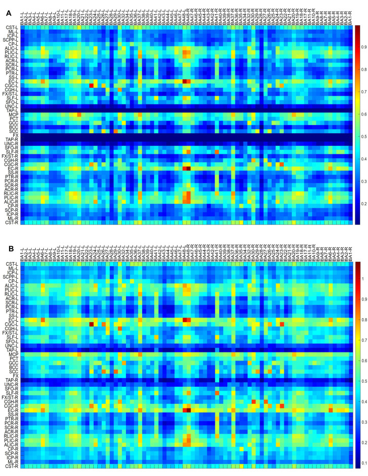
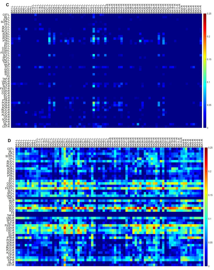
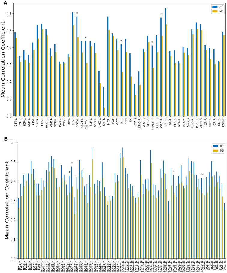
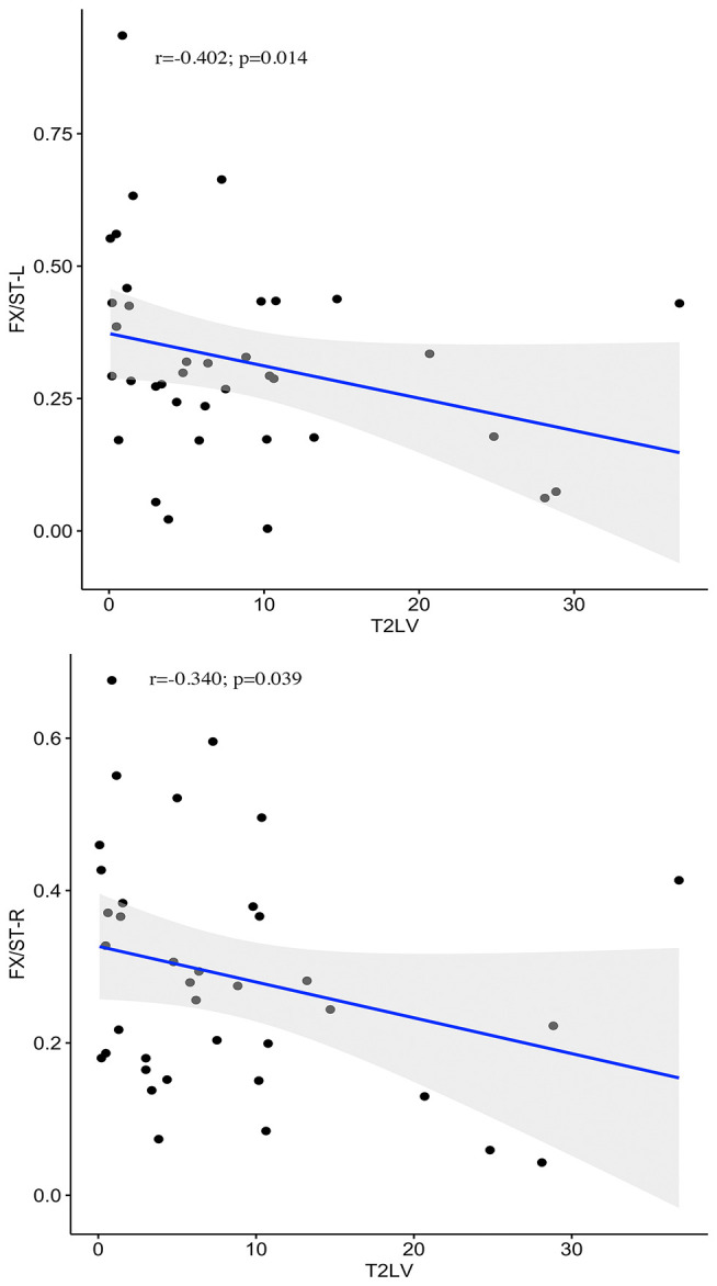
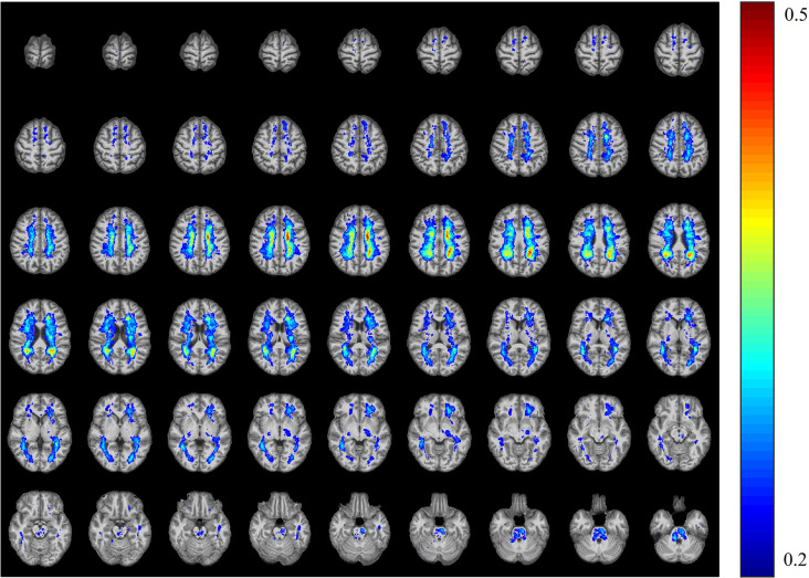
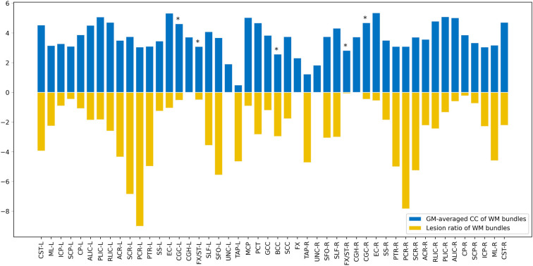
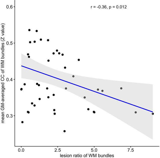
Background: Functional magnetic resonance imaging (fMRI) has been widely used to assess neural activity changes in gray matter (GM) in patients with multiple sclerosis (MS); however, brain function alterations in white matter (WM) relatively remain under-explored. Purpose: This work aims to identify the functional connectivity in both the WM and the GM of patients with MS using fMRI and the correlations between these functional changes and cumulative disability as well as the lesion ratio. Materials and Methods: For this retrospective study, 37 patients with clinically definite MS and 43 age-matched healthy controls were included between 2010 and 2014. Resting-state fMRI was performed. The WFU Pick and JHU Eve atlases were used to define 82 GM and 48 WM regions in common spaces, respectively. The time courses of blood oxygen level-dependent (BOLD) signals were averaged over each GM or WM region. The averaged time courses for each pair of GM and WM regions were correlated. All 82 × 48 correlations for each subject formed a functional correlation matrix. Results: Compared with the healthy controls, the MS patients had a decreased temporal correlation between the WM and the GM regions. Five WM bundles and four GM regions had significantly decreased mean correlation coefficients (CCs). More specifically, the WM functional alterations were negatively correlated with the lesion volume in the bilateral fornix, and the mean GM-averaged CCs of the WM bundles were inversely correlated with the lesion ratio (r = -0.36, P = 0.012). No significant correlation was found between WM functional alterations and the paced auditory serial addition test score, Expanded Disease Severity Scale score, and Multiple Sclerosis Severity Score (MSSS) in MS. Conclusions: These findings highlight current gaps in our knowledge of the WM functional alterations in patients with MS and may link WM function with pathological mechanisms.
Keywords: MRI; functional connectivity; multiple sclerosis; resting-state; white matter.
Copyright © 2020 Huang, Li, Li, Yang, Xin, Qi, Liu, Dong, Li, Ding and Lu.
Conflict of interest statement
The authors declare that the research was conducted in the absence of any commercial or financial relationships that could be construed as a potential conflict of interest.
Figures
Similar articles
-
Disrupted White Matter Functional Connectivity With the Cerebral Cortex in Migraine Patients.Front Neurosci. 2022 Jan 13;15:799854. doi: 10.3389/fnins.2021.799854. eCollection 2021. Front Neurosci. 2022. PMID: 35095401 Free PMC article.
-
Detection and quantification of regional cortical gray matter damage in multiple sclerosis utilizing gradient echo MRI.Neuroimage Clin. 2015 Aug 18;9:164-75. doi: 10.1016/j.nicl.2015.08.003. eCollection 2015. Neuroimage Clin. 2015. PMID: 27330979 Free PMC article.
-
Characterization of gray-matter multiple sclerosis lesions using double inversion recovery, diffusion, contrast-enhanced, and volumetric MRI.Mult Scler Relat Disord. 2019 Jun;31:74-81. doi: 10.1016/j.msard.2019.03.021. Epub 2019 Mar 29. Mult Scler Relat Disord. 2019. PMID: 30951968
-
Functional MRI and resting state connectivity in white matter - a mini-review.Magn Reson Imaging. 2019 Nov;63:1-11. doi: 10.1016/j.mri.2019.07.017. Epub 2019 Jul 31. Magn Reson Imaging. 2019. PMID: 31376477 Free PMC article. Review.
-
Gray matters in multiple sclerosis: cognitive impairment and structural MRI.Mult Scler Int. 2014;2014:609694. doi: 10.1155/2014/609694. Epub 2014 Jan 22. Mult Scler Int. 2014. PMID: 24587905 Free PMC article. Review.
Cited by
-
Changes in white matter functional networks across late adulthood.Front Aging Neurosci. 2023 Jun 30;15:1204301. doi: 10.3389/fnagi.2023.1204301. eCollection 2023. Front Aging Neurosci. 2023. PMID: 37455933 Free PMC article.
-
Overview of the Current Pathophysiology of Fatigue in Multiple Sclerosis, Its Diagnosis and Treatment Options - Review Article.Neuropsychiatr Dis Treat. 2023 Nov 20;19:2485-2497. doi: 10.2147/NDT.S429862. eCollection 2023. Neuropsychiatr Dis Treat. 2023. PMID: 38029042 Free PMC article. Review.
-
Disrupted White Matter Functional Connectivity With the Cerebral Cortex in Migraine Patients.Front Neurosci. 2022 Jan 13;15:799854. doi: 10.3389/fnins.2021.799854. eCollection 2021. Front Neurosci. 2022. PMID: 35095401 Free PMC article.
-
Quantification of mediation effects of white matter functional characteristics on cognitive decline in aging.Cereb Cortex. 2024 Mar 1;34(3):bhae114. doi: 10.1093/cercor/bhae114. Cereb Cortex. 2024. PMID: 38517178 Free PMC article.
-
Dynamic variations of resting-state BOLD signal spectra in white matter.Neuroimage. 2022 Apr 15;250:118972. doi: 10.1016/j.neuroimage.2022.118972. Epub 2022 Feb 4. Neuroimage. 2022. PMID: 35131432 Free PMC article.
References
-
- Bodini B., Khaleeli Z., Cercignani M., Miller D. H., Thompson A. J., Ciccarelli O. (2009). Exploring the relationship between white matter and gray matter damage in early primary progressive multiple sclerosis: an in vivo study with TBSS and VBM. Hum. Brain Mapp. 30, 2852–2861. 10.1002/hbm.20713 - DOI - PMC - PubMed
LinkOut - more resources
Full Text Sources