. 2020 Dec 4:12:596551.
doi: 10.3389/fnagi.2020.596551. eCollection 2020.

Neural Generators Underlying Temporal Envelope Processing Show Altered Responses and Hemispheric Asymmetry Across Age

Affiliations

Neural Generators Underlying Temporal Envelope Processing Show Altered Responses and Hemispheric Asymmetry Across Age

Ehsan Darestani Farahani et al. Front Aging Neurosci. .

Abstract

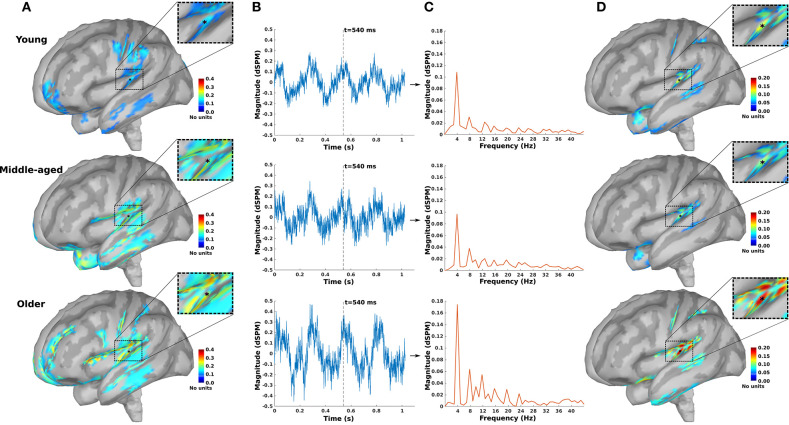
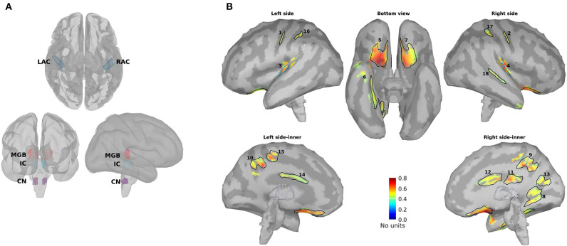
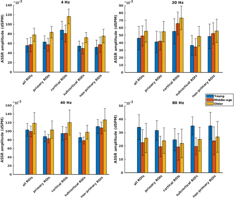
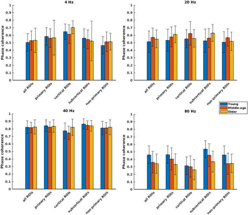
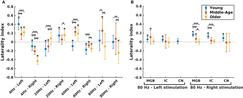
Speech understanding problems are highly prevalent in the aging population, even when hearing sensitivity is clinically normal. These difficulties are attributed to changes in central temporal processing with age and can potentially be captured by age-related changes in neural generators. The aim of this study is to investigate age-related changes in a wide range of neural generators during temporal processing in middle-aged and older persons with normal audiometric thresholds. A minimum-norm imaging technique is employed to reconstruct cortical and subcortical neural generators of temporal processing for different acoustic modulations. The results indicate that for relatively slow modulations (<50 Hz), the response strength of neural sources is higher in older adults than in younger ones, while the phase-locking does not change. For faster modulations (80 Hz), both the response strength and the phase-locking of neural sources are reduced in older adults compared to younger ones. These age-related changes in temporal envelope processing of slow and fast acoustic modulations are possibly due to loss of functional inhibition, which is accompanied by aging. Both cortical (primary and non-primary) and subcortical neural generators demonstrate similar age-related changes in response strength and phase-locking. Hemispheric asymmetry is also altered in older adults compared to younger ones. Alterations depend on the modulation frequency and side of stimulation. The current findings at source level could have important implications for the understanding of age-related changes in auditory temporal processing and for developing advanced rehabilitation strategies to address speech understanding difficulties in the aging population.

Keywords: ASSR; EEG; aging; auditory steady-state response; auditory temporal processing; neural generators.


Conflict of interest statement

The authors declare that the research was conducted in the absence of any commercial or financial relationships that could be construed as a potential conflict of interest.

Figures

Figure 1
Figure 1
The auditory steady-state response (ASSR) map in response to 4 Hz amplitude-modulated (AM) stimuli presented to the right ear across age. (A) Reconstructed source map at 540 ms using dynamic statistical parametric mapping (dSPM) and enlarged view of a sample dipole located in the auditory cortex [−35, −28, 16, xyz in Montreal Neurological Institute (MNI) coordinates]. The map illustrates the absolute values of activity, and the color bar indicates the magnitude of activity (no unit because of normalization, which is performed within the dSPM algorithm). (B) Time-series of the sample dipole (original values with length of one epoch) for the three age cohorts. The vertical dashed line indicates the time point of 540 ms. (C) The frequency spectrum of the sample dipole for the three age cohorts. (D) The generated ASSR map for the three age cohorts. The color bar indicates the ASSR amplitude.
Figure 2
Figure 2
Primary and non-primary regions of interest (ROIs). (A) The primary ROIs are located bilaterally in the auditory cortex (LAC, RAC), the medial geniculate body (LMGB, RMGB), inferior colliculus (LIC, RIC), and cochlear nucleus (LCN, RCN). (B) The non-primary ROIs were based on the averaged normalized signal-to-noise ratio (SNR) maps of all experimental conditions [young, middle aged, and older cohort, 4, 20, 40, and 80 Hz amplitude-modulated (AM) stimuli presented to the left and the right ears] and the obtained ROIs. The anatomical labels of the primary and the non-primary ROIs are listed in Table 1.
Figure 3
Figure 3
Auditory steady-state response (ASSR) amplitudes of different categories of sources (Table 1) regardless of side of stimulation across age and across modulation frequency. The bars indicate the weighted average of amplitudes (number of subjects as weights), and error bars represent the pooled standard deviations (Cohen, 1988). Forty-eight of 60 comparisons showed significant difference (Table 2).
Figure 4
Figure 4
Phase coherence of different categories of sources (Table 1), regardless of side of stimulation across age and across modulation frequency. The bars indicate the weighted average of phase coherence (number of subjects as weights), and error bars represent the pooled standard deviations (Cohen, 1988). Twenty-six of 60 comparisons showed a significant difference (Table 2).
Figure 5
Figure 5
Hemispheric lateralization for different age groups. (A) The laterality indexes (LIs) for auditory cortex (AC) across age (indicated by different colors) in different experimental conditions (four modulation frequencies presented to the left or right ear). (B) The LIs for the subcortical sources [the medial geniculate body (MGB), the inferior colliculus (IC), the cochlear nucleus (CN)] across age (indicated by different colors) in response to 80 Hz amplitude-modulated (AM) stimuli presented to the left or right ear. LI for CN (left stimulation) was not calculated because of non-significant auditory steady-state response (ASSR) amplitude in one side. The LIs were obtained based on the 17.7 (±0.4), 17.6 (±0.7), and 14.9 (±1) participants in the young, middle-aged, and older cohorts. The error bars illustrate the standard deviations estimated using the jackknife method (Efron and Stein, 1981). The significant left/right lateralization was indicated by an asterisk (*) on top of the error bar. Comparisons across age: *p ≤ 0.05; **p ≤ 0.01; ***p ≤ 0.001.

Similar articles

Cited by

References

    1. Anderson S., Parbery-Clark A., White-Schwoch T., Kraus N. (2012). Aging affects neural precision of speech encoding. J. Neurosci. 32, 14156–14164. 10.1523/JNEUROSCI.2176-12.2012 - DOI - PMC - PubMed
    1. Benjamini Y., Hochberg Y. (1995). Controlling the false discovery rate: a practical and powerful approach to multiple testing. J. R. Stat. Soc. Ser. B 57, 289–300. 10.1111/j.2517-6161.1995.tb02031.x - DOI
    1. Berlingeri M., Danelli L., Bottini G., Sberna M., Paulesu E. (2013). Reassessing the HAROLD model: is the hemispheric asymmetry reduction in older adults a special case of compensatory-related utilisation of neural circuits? Exp. Brain Res. 224, 393–410. 10.1007/s00221-012-3319-x - DOI - PubMed
    1. Boettcher F. A., Poth E. A., Mills J. H., Dubno J. R. (2001). The amplitude-modulation following response in young and aged human subjects. Hear. Res. 153, 32–42. 10.1016/S0378-5955(00)00255-0 - DOI - PubMed
    1. Bradley A., Yao J., Dewald J., Richter C. P. (2016). Evaluation of electroencephalography source localization algorithms with multiple cortical sources. PLoS ONE 11:e0147266. 10.1371/journal.pone.0147266 - DOI - PMC - PubMed

LinkOut - more resources