

The Expanding Cell Diversity of the Brain Vasculature
- PMID: 33343397
- PMCID: PMC7744630
- DOI: 10.3389/fphys.2020.600767
The Expanding Cell Diversity of the Brain Vasculature
Abstract
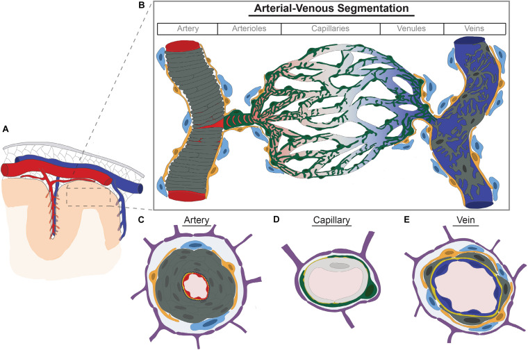
The cerebrovasculature is essential to brain health and is tasked with ensuring adequate delivery of oxygen and metabolic precursors to ensure normal neurologic function. This is coordinated through a dynamic, multi-directional cellular interplay between vascular, neuronal, and glial cells. Molecular exchanges across the blood-brain barrier or the close matching of regional blood flow with brain activation are not uniformly assigned to arteries, capillaries, and veins. Evidence has supported functional segmentation of the brain vasculature. This is achieved in part through morphologic or transcriptional heterogeneity of brain vascular cells-including endothelium, pericytes, and vascular smooth muscle. Advances with single cell genomic technologies have shown increasing cell complexity of the brain vasculature identifying previously unknown cell types and further subclassifying transcriptional diversity in cardinal vascular cell types. Cell-type specific molecular transitions or zonations have been identified. In this review, we summarize emerging evidence for the expanding vascular cell diversity in the brain and how this may provide a cellular basis for functional segmentation along the arterial-venous axis.
Keywords: astrocytes; blood brain barrier; endothelial cells; neurovascular unit; pericytes and vascular smooth muscle cells; perivascular fibroblasts; perivascular macrophages; single cell sequencing.
Copyright © 2020 Ross, Kim, Allen, Crouch, Narsinh, Cooke, Abla, Nowakowski and Winkler.
Conflict of interest statement
The authors declare that the research was conducted in the absence of any commercial or financial relationships that could be construed as a potential conflict of interest.
Figures
Similar articles
-
Defective vascular signaling & prospective therapeutic targets in brain arteriovenous malformations.Neurochem Int. 2019 Jun;126:126-138. doi: 10.1016/j.neuint.2019.03.002. Epub 2019 Mar 8. Neurochem Int. 2019. PMID: 30858016 Review.
-
Public Volume Electron Microscopy Data: An Essential Resource to Study the Brain Microvasculature.Front Cell Dev Biol. 2022 Apr 5;10:849469. doi: 10.3389/fcell.2022.849469. eCollection 2022. Front Cell Dev Biol. 2022. PMID: 35450291 Free PMC article.
-
Brain macrophages: on the role of pericytes and perivascular cells.Brain Res Brain Res Rev. 1999 Dec;31(1):42-57. doi: 10.1016/s0165-0173(99)00024-7. Brain Res Brain Res Rev. 1999. PMID: 10611494 Review.
-
Location Matters: Navigating Regional Heterogeneity of the Neurovascular Unit.Front Cell Neurosci. 2021 Jun 30;15:696540. doi: 10.3389/fncel.2021.696540. eCollection 2021. Front Cell Neurosci. 2021. PMID: 34276312 Free PMC article. Review.
-
A molecular atlas of cell types and zonation in the brain vasculature.Nature. 2018 Feb 22;554(7693):475-480. doi: 10.1038/nature25739. Epub 2018 Feb 14. Nature. 2018. PMID: 29443965
Cited by
-
Role of TRP ion channels in cerebral circulation and neurovascular communication.Neurosci Lett. 2021 Nov 20;765:136258. doi: 10.1016/j.neulet.2021.136258. Epub 2021 Sep 22. Neurosci Lett. 2021. PMID: 34560190 Free PMC article. Review.
-
Single-cell RNA sequencing reveals dysregulation of spinal cord cell types in a severe spinal muscular atrophy mouse model.PLoS Genet. 2022 Sep 8;18(9):e1010392. doi: 10.1371/journal.pgen.1010392. eCollection 2022 Sep. PLoS Genet. 2022. PMID: 36074806 Free PMC article.
-
Beyond the neuron: Role of non-neuronal cells in stress disorders.Neuron. 2022 Apr 6;110(7):1116-1138. doi: 10.1016/j.neuron.2022.01.033. Epub 2022 Feb 18. Neuron. 2022. PMID: 35182484 Free PMC article. Review.
-
Molecular Heterogeneity of the Brain Endothelium.Curr Issues Mol Biol. 2023 Apr 16;45(4):3462-3478. doi: 10.3390/cimb45040227. Curr Issues Mol Biol. 2023. PMID: 37185751 Free PMC article.
-
Neuronal activity drives IGF2 expression from pericytes to form long-term memory.Neuron. 2023 Dec 6;111(23):3819-3836.e8. doi: 10.1016/j.neuron.2023.08.030. Epub 2023 Oct 2. Neuron. 2023. PMID: 37788670 Free PMC article.