Review
. 2020 Dec 3:11:591741.
doi: 10.3389/fimmu.2020.591741. eCollection 2020.

Angiogenesis in Lymph Nodes Is a Critical Regulator of Immune Response and Lymphoma Growth

Affiliations
Review

Angiogenesis in Lymph Nodes Is a Critical Regulator of Immune Response and Lymphoma Growth

Lutz Menzel et al. Front Immunol. .

Abstract

Tumor-induced remodeling of the microenvironment in lymph nodes (LNs) includes the formation of blood vessels, which goes beyond the regulation of metabolism, and shaping a survival niche for tumor cells. In contrast to solid tumors, which primarily rely on neo-angiogenesis, hematopoietic malignancies usually grow within pre-vascularized autochthonous niches in secondary lymphatic organs or the bone marrow. The mechanisms of vascular remodeling in expanding LNs during infection-induced responses have been studied in more detail; in contrast, insights into the conditions of lymphoma growth and lodging remain enigmatic. Based on previous murine studies and clinical trials in human, we conclude that there is not a universal LN-specific angiogenic program applicable. Instead, signaling pathways that are tightly connected to autochthonous and infiltrating cell types contribute variably to LN vascular expansion. Inflammation related angiogenesis within LNs relies on dendritic cell derived pro-inflammatory cytokines stimulating vascular endothelial growth factor-A (VEGF-A) expression in fibroblastic reticular cells, which in turn triggers vessel growth. In high-grade B cell lymphoma, angiogenesis correlates with poor prognosis. Lymphoma cells immigrate and grow in LNs and provide pro-angiogenic growth factors themselves. In contrast to infectious stimuli that impact on LN vasculature, they do not trigger the typical inflammatory and hypoxia-related stroma-remodeling cascade. Blood vessels in LNs are unique in selective recruitment of lymphocytes via high endothelial venules (HEVs). The dissemination routes of neoplastic lymphocytes are usually disease stage dependent. Early seeding via the blood stream requires the expression of the homeostatic chemokine receptor CCR7 and of L-selectin, both cooperate to facilitate transmigration of tumor and also of protective tumor-reactive lymphocytes via HEV structures. In this view, the HEV route is not only relevant for lymphoma cell homing, but also for a continuous immunosurveillance. We envision that HEV functional and structural alterations during lymphomagenesis are not only key to vascular remodeling, but also impact on tumor cell accessibility when targeted by T cell-mediated immunotherapies.

Keywords: B cell malignancy; angiogenesis; high endothelial venule; lymph node; lymphocyte trafficking; lymphoma; reactive endothelium; tumor microenvironment.


Conflict of interest statement

The authors declare that the research was conducted in the absence of any commercial or financial relationships that could be construed as a potential conflict of interest.

Figures
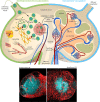
Figure 1
Figure 1
Lymph node vascularization in development and under homeostatic conditions. The LN compartments during LN development (left) and homeostatic conditions (right). Left: Lymphoid organogenesis is driven by recruitment of Lymphoid tissue-inducer (LTi) cells that stimulate lymphoid-organizer (LTo) cells via lymphotoxin (LT) α1β2 - LT β receptor signaling, which secrete LTi-recruiting CXCL13 in turn. LTi recruitment from the blood circulation and the afferent lymphatics accumulates LTi cells within the LN anlagen resulting in a self-amplifying process of LN development. α4β7 integrin-expressing LTi recruitment and extravasation utilizes the mucosal vascular addressin cell adhesion molecule-1 (MadCAM-1) on the luminal surface of blood vessels. MadCAM-1 switches to peripheral node addressin (PNAd) expression during differentiation of mature high endothelial venules (HEVs) within peripheral LNs. The formation of the blood vessel network comprises sprouting and branching of expanding blood vessels driven by retinoic acid (RA) stimulation of the RA receptor (RAR) on blood endothelial cells (BECs). Right: The blood circulation enters the LN during homeostatic conditions via the feeding arteriole at the LN hilum, proceeds along the medullary cord and branches into metarterioles that feed the capillary networks around the medulla and at the subcapsular sinus. HEVs are post-capillary venules with a characteristically enlarged vessel diameter. The venous backflow leaves the LN in a bundle of venules at the hilum. Bottom: Representative histochemistry sections (vessels: Cadherin5 fluoresent_reporter, red) of murine LNs during homeostasis and during progression of a murine high-grade B cell lymphoma.
Figure 2
Figure 2
Lymphoma induced angiogenesis in LNs and participating immune cells. Top: The LN compartments represented under homeostatic conditions (left) and lymphoma-activated angiogenesis (right). Lymphoma growth is characterized by a strong LN volume expansion and blood vasculature growth. Remodeling of the stromal infrastructure involves an increase of the microvessel density (MVD), as effectuated by direct angiogenic stimulation through lymphoma B cells cells, but concomitantly also through reciprocal crosstalk of cells in the TME and recruited immune cells. Notably, the initiation of the angiogenic switch in lymphoma is independent from hypoxia-induced HIF1α pathway activation. Tumor polarized DCs (CEBP/βhigh) control the HEV differentiation status via LTα1β2 and LIGHT presentation; they release IL-1β and hereby take part in the blood vessel growth by inducing VEGF-A expression in FRCs. They also secrete the angiogenic factors VEGF-A and FGF2. B cells express LTα1β2, which exerts minor effects on HEVs, but a predominating stimulatory effect on FRCs. Expression of the chemokines CCL2, CXCL12, and MIF recruits additional immune cells into the LN. Regulatory T cells (Tregs), myeloid-derived suppressor cells (MDSCs), M2-polarized macrophages, neutrophils and mast cells are capable of producing the pro-angiogenic factors VEGF-A, VEGF-B, VEGF-C, MMP9, IL-8, IL-10, TGFβ, and FGF1/2. Bottom left: HEVs express PNAd, CCL21, and ICAM1 and thereby constitute the transmigration routes for lymphocytes under homeostatic conditions. Interaction of CD62L, CCR7, and LFA-1 on naïve lymphocytes with these HEV-associated surface receptors and chemokines initiates lymphocyte rolling, HEV wall adhesion and eventually, transmigration into the LN parenchyma. Bottom middle: Inflammatory vessels in reactive LNs recruit activated lymphocytes by CXCL9 secretion and replace the homeostatic receptors on endothelial cells with CD62P, CD63E, and VCAM1 that are interaction partners of leukocyte-expressed CD44, PSGL1, and VLA4. Bottom right: The lymphoma induced expansion of the blood vessel network favors the assembly of smaller anergic endothelium that is insufficiently equipped for lymphocyte extravasation.
Figure 3
Figure 3
Therapeutic strategies to induce vessel normalization and revert endothelial anergy in B-NHL. (A) Anti-angiogenesis therapy tageted at VEGFR-2 or VEGFR-3 can restore a normalized vessel network. (B) Targeting of the LTβR with LTα1β2 and LIGHT expressing DCs, agonistic antibodies or recombinant factors potentially circumvents impaired lymphocyte homing by establishing or stabilizing HEV integrity within the lymphoma TME (338). (C) Vessel anergy can be changed by a targeted conversion of the endothelium toward a reactive endothelium using inflammatory cytokines, which might be site directed to avoid unintended systemic effects. Normalization of aberrant vessels and activation of the endothelium can also be achieved by locally applied low-dose gamma irradiation (339). Reactive endothelium within LNs is a prerequisite for an effective infiltration of effector T cells during cellular immunotherapy.

References

    1. Krishnamurty AT, Turley SJ. Lymph node stromal cells: cartographers of the immune system. Nat Immunol (2020). 10.1038/s41590-020-0635-3 - DOI - PubMed
    1. Till KJ, Coupland SE, Pettitt AR. Motility and trafficking in B-cell non-Hodgkin’s lymphoma (Review). Int J Oncol (2014). 10.3892/ijo.2014.2395 - DOI - PubMed
    1. Wang L, Qin W, Huo YJ, Li X, Shi Q, Rasko JEJ, et al. Advances in targeted therapy for malignant lymphoma. Signal Transduct Target Ther (2020). 10.1038/s41392-020-0113-2 - DOI - PMC - PubMed
    1. Maman S, Witz IP. A history of exploring cancer in context. Nat Rev Cancer (2018). 10.1038/s41568-018-0006-7 - DOI - PubMed
    1. Hanahan D, Weinberg RA. Hallmarks of cancer: The next generation. Cell (2011). 10.1016/j.cell.2011.02.013 - DOI - PubMed

Publication types