. 2020 Mar;11(1):421-439.
doi: 10.1146/annurev-conmatphys-031218-013231. Epub 2019 Dec 6.

The Actin Cytoskeleton as an Active Adaptive Material

Affiliations

The Actin Cytoskeleton as an Active Adaptive Material

Shiladitya Banerjee et al. Annu Rev Condens Matter Phys. 2020 Mar.

Abstract

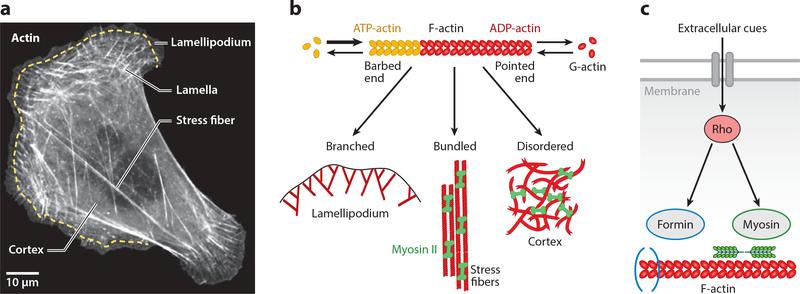
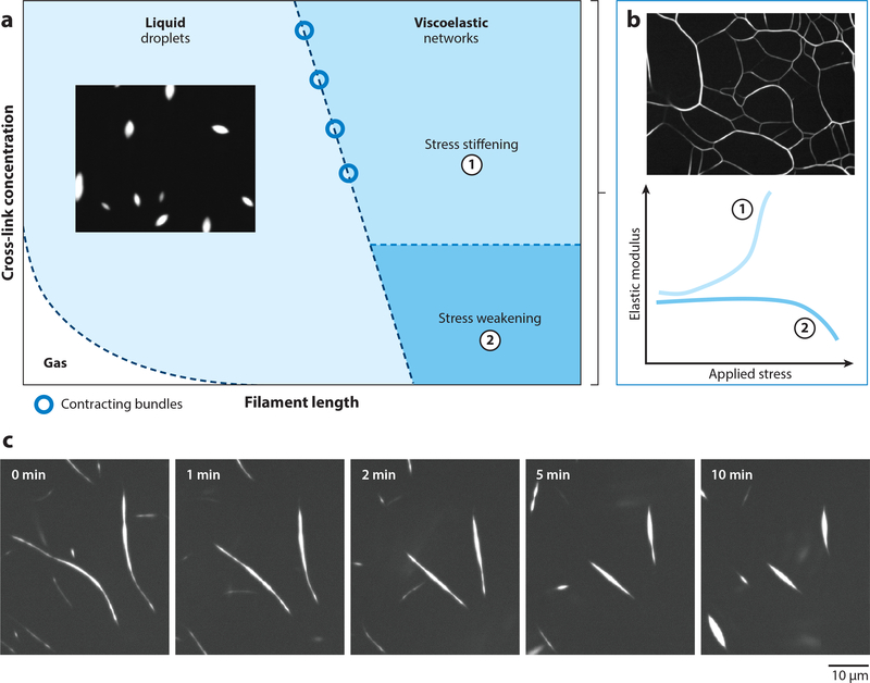
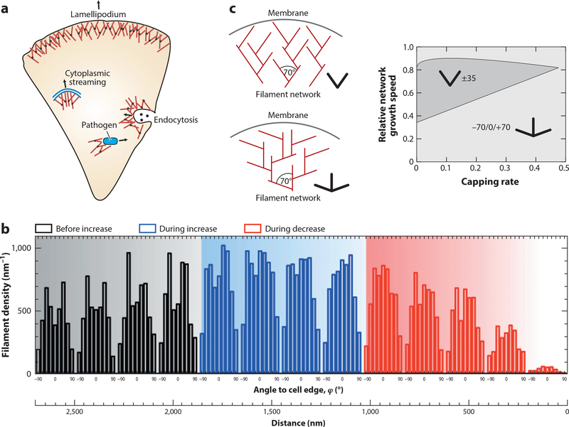
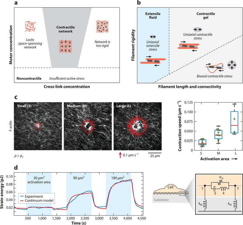
Actin is the main protein used by biological cells to adapt their structure and mechanics to their needs. Cellular adaptation is made possible by molecular processes that strongly depend on mechanics. The actin cytoskeleton is also an active material that continuously consumes energy. This allows for dynamical processes that are possible only out of equilibrium and opens up the possibility for multiple layers of control that have evolved around this single protein.Here we discuss the actin cytoskeleton from the viewpoint of physics as an active adaptive material that can build structures superior to man-made soft matter systems. Not only can actin be used to build different network architectures on demand and in an adaptive manner, but it also exhibits the dynamical properties of feedback systems, like excitability, bistability, or oscillations. Therefore, it is a prime example of how biology couples physical structure and information flow and a role model for biology-inspired metamaterials.

Keywords: active matter; cell mechanics; feedback control; metamaterial; nonequilibrium physics; soft matter.


Figures

Figure 1
Figure 1
Actin networks organize in distinct architectures and modules in cells. (a) Actin organization in a U2OS cell, visualized by fluorescent actin. The actin cytoskeleton organizes into diverse superstructures in cells, including branched networks in the lamellipodium at the cell front, contractile transverse arcs in the lamella behind the lamellipodium, cross-linked and contractile meshworks in the cortex, and stress fibers stretching toward the cell rear. Scale bar represents 10 μm. Panel adapted from Reference . (b) Actin is a living polymer that utilizes energy from ATP-hydrolysis to assemble monomers at the barbed end and to disassemble them from the pointed end. By associating with specific binding partners, actin can assemble the diverse architectures seen in panel a.(c) Flow of information toward actin. Extracellular cues are integrated by membrane receptors to activate signaling pathways, including those that regulate the assembly of actin structures. As an example, here we show the Rho pathway that synchronizes the assembly of the actomyosin system through formin-mediated actin polymerization and myosin II–driven contractility.
Figure 2
Figure 2
Materials properties of cross-linked actin networks. (a) State diagram of cross-linked actin networks showing the tunability of mechanical properties by varying cross-link concentration and filament length. When filament lengths are sufficiently long for entanglements, F-actin kinetically arrests to form viscoelastic networks. Short F-actin with sufficiently high cross-linking concentration forms liquid droplets. Gas-like phase appears when both filament length and cross-linking concentration are small. (b) Tunability of elastic modulus of cross-linked F-actin in the viscoelastic phase. F-actin networks stress-stiffen when densely cross-linked and stress-weaken for low cross-linking concentrations. (c) At the boundary between viscoelastic and fluid phases, cross-linked F-actin forms long bundles that contract via surface-tension-like forces. Figure adapted from Reference .
Figure 3
Figure 3
Active growth of F-actin networks. (a) In cells, dendritic actin networks generated by the branching agent Arp2/3 are used to create pushing forces on the objects that nucleate them, for example, on the plasma membrane during migration, on endocytosing vesicles that have to be pushed inside the cell during cytoplasmic streaming and when pathogens push themselves through the cytoplasm. (b) When membrane tension and thus mechanical load on the lamellipodium is first increased and then decreased by micropipette aspiration of migrating keratocytes, filament density first increases and then decreases. At the same time, the orientation distribution changes from two symmetric peaks at ±35° to one peak at 0°. Panel adapted from Reference with permission. (c) These results agree well with the theoretical predictions that the classical ±35° structure (slingshot) competes with a +70/0/−70° structure (trident) depending on the growth kinetics determined by external load. Panel adapted from Reference .
Figure 4
Figure 4
Contractile force propagation by actomyosin networks. (a) Phase diagram showing the dependence of macroscopic contractility of F-actin networks on the concentration of cross-linker and myosin II motors. The network contracts for high-enough motor concentration and at intermediate concentration of cross-linkers. Low cross-linking does not yield space-spanning networks, whereas high cross-linking makes the network too rigid to contract. (b) Phase diagram showing the mechanisms for actomyosin contractility by varying network connectivity (cross-link concentration or filament length) or filament rigidity, at high-enough motor concentration. Rigid filaments with low cross-linking yield extensile fluid networks. Panel adapted from Reference . (c)(Left) Actomyosin network contraction in vitro for myosin activation regions of varying areas, with red vectors showing the velocity of contraction at the activation boundary. (Right) Contraction velocity of actomyosin scales with myosin activation area. Panel adapted from Reference . (d)(Left) Data from live cell experiments showing the dependence of contractile strain energy (measured by traction force microscopy) on the area of RhoA activation by optogenetics. (Right) Schematic of a continuum mechanical model for a contractile adherent cell. Panel adapted from Reference .
Figure 5
Figure 5
Feedback control systems in the actin cytoskeleton. (a) Rho-GTPases are localized to the cell membrane and cycle between an active GTP-bound state and an inactive GDP-bound state. The switching rates are determined by a large range of regulators (GEFs for activation and GAPs for inactivation). (b) A simplified diagram of the cross talks and feedback loops in the actin cytoskeleton controlled by the small GTPases of the Rho family. (c) Negative feedback control of protrusive activity in the lamellipodium controlled by the Rac pathway. (d) Positive feedback control of stress fiber assembly and contraction, downstream of the Rho pathway. (e) The actin cortex exhibits a mixture of contractile and protrusive activities downstream of the Rho and Rac pathways. Abbreviations: GAP, GTPase-activating protein; GDP, guanosine diphosphate; GEF, guanine nucleotide exchange factor; GTP, guanosine triphosphate.
Figure 6
Figure 6
Excitability, bistability, and traveling waves in the actin cortex. (a) Schematic of a typical activator-inhibitor feedback system. (b,c) Spatial profiles of activator (red) and inhibitor (blue) concentrations in a traveling excitable wave (b) and in a static structure (c). Panel adapted from Reference . (d) RhoA-actin feedback system in the cortex as an activator-inhibitor system. (e,f) Excitability in the vicinity of the cytokinetic furrow (e) and stable coexistence of high RhoA activity and F-actin concentration in the middle of the furrow (f). GEF concentration is shown in red shading. Phase trajectories are shown for F-actin nullcline (blue curve), Rho nullcline (red curve), an excitable trajectory (dashed curve), and resting steady state of the cortex (green solid circle). Panels e and f adapted from Reference . (g) Phase plot in RhoA-actin system illustrating that diffusion can move the F-actin concentration across the threshold for transition to the high-activity state. (h,i) Spatial coupling in the activator-inhibitor system can give rise to excitable (h)orbistable(i) waves. Red shading represents activity. Abbreviations: GAP, GTPase-activating protein; GDP, guanosine diphosphate; GEF, guanine nucleotide exchange factor; GTP, guanosine triphosphate.

References

    1. Alberts B, Johnson A, Lewis J, Morgan D, Raff M, et al. 2014. Molecular Biology of the Cell. New York: Norton & Co; 6th rev. ed.
    1. Phillips R, Kondev J, Theriot J, Garcia H. 2012. Physical Biology of the Cell. New York: Taylor & Francis; 2nd ed.
    1. Pollard TD, Cooper JA. 2009. Science 326(5957):1208–12 - PMC - PubMed
    1. Jockusch BM, ed. 2017. The Actin Cytoskeleton. Berlin: Springer
    1. Stricker J, Falzone T, Gardel ML. 2010. J. Biomech. 43(1):9–14 - PMC - PubMed

LinkOut - more resources