. 2020 Nov 10;12(6):plaa057.
doi: 10.1093/aobpla/plaa057. eCollection 2020 Dec.

Homology and functions of inner staminodes in Anaxagorea javanica (Annonaceae)

Affiliations

Homology and functions of inner staminodes in Anaxagorea javanica (Annonaceae)

Bingxin Li et al. AoB Plants. .

Abstract

Inner staminodes are widespread in Magnoliales and present in Anaxagorea and Xylopia, but were lost in the other genera of Annonaceae and have no counterparts in derived angiosperms. The coexistence of normal stamens, modified stamens and inner staminodes in Anaxagorea javanica is essential to understand the homology and pollination function of the inner staminodes. Anaxagorea javanica was subjected to an anatomical study by light and scanning electron microscopy, and the chemistry of secretions was evaluated by an amino acid analyser. Inner staminodes have a secretory apex, but do not have thecae. They bend towards either tepals or carpels at different floral stages, and function as a physical barrier preventing autogamy and promoting outcrossing. At the pistillate phase, the exudates from the inner staminodes have high concentration of amino acid, and provide attraction to pollinating insects; while abundant proline was only detected in stigmas exudates, and supply for pollen germination. Modified stamens have a secretory apex and one or two thecae, which are as long as or shorter than that of the normal stamens. As transitional structures, modified stamens imply a possible degeneration progress from normal stamens to inner staminodes: generating a secretory apex first, shortening of the thecae length next and then followed by the loss of thecae. The presence of modified stamens together with the floral vasculature and ontogeny imply that the inner staminodes are homologous with stamens.

Keywords: Anaxagorea javanica; Annonaceae; homology; inner staminode; modified stamen; pollination function.


Figures

Figure 1.
Figure 1.
Floral phenology of A. javanica during anthesis. (A) The petals of A. javanica are slightly opened. (B) Nitidulid beetles visiting gaps between petals at the pistillate phase. (C) The inner staminodes bend towards the stamens and away from the pistils at the pistillate phase. (D) Flower of A. javanica at the interim phase. (E) The inner staminodes start bending towards pistils at the interim phase. (F) Flower of A. javanica at the beginning of the staminate phase. (G) The inner staminodes are curved over the stigmas at the staminate phase. (H) The petals are widely opened at the end of staminate phase; pollinator (arrow) enters a gap between the petals. (I) There are several Colopterus spp. (Nitidulidae) dusted with pollen inside of the flower at the end of the staminate phase. C, carpel; S, stamen; St, staminode.
Figure 2.
Figure 2.
Timing of anthesis of A. javanica. (1) Flowers spreading their petals. (2) Initiation of scent production. (3) Initiation of staminate stage. (4) Arrival of pollinators. (5) The end of the staminate stage.
Figure 3.
Figure 3.
Microphotographs of the two putatively most important pollinators. (A, B, E–G) Colopterus spp. (Nitidulidae). (A) Dorsal view. (B) Ventral view. (E–G) Detail of pollen sticking to the abdomen and legs of Colopterus spp. (C, D, H–J) Epuraea spp. (Nitidulidae). (C) Dorsal view. (D) Ventral view. (H–J) Detail of pollen sticking to the abdomen and legs of Epuraea spp. Scale bars: A–D = 250 μm; E–J = 50 μm.
Figure 4.
Figure 4.
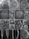
The morphology of modified stamens and pollen grains from the normal stamens and the modified ones. (A–C) Modified stamens located between the normal stamens and the inner staminodes. (A) Flowers with one modified stamen possessing a secretory apex and two shorter thecae than in the normal stamens. (B, C) Flowers with two adjacent modified stamens covered by secretory structures at the apex: one with two thecae of the usual length or shorter thecae, the other with one shorter theca. (D, E) Modified stamens with two shorter thecae than in the normal stamens. (F) Modified stamen with one shorter theca. (G–I) The apex of modified stamens covered by secretory structures. (J) Longitudinal microtome sections of the apex of inner staminodes, showing one basal cell, three stalk cells and one head cell. (K, M) Pollen grains from a normal stamen. (L, N) Pollen grains from a modified stamen. (O) Germinating pollen from a normal stamen producing a pollen tube. (P) Germinating pollen from a modified stamen producing a pollen tube. BC, basal cell; C, carpel; HC, head cell; S, stamen; SC, stalk cell; MS, modified stamen; St, staminode. Scale bars: D–F = 500 μm; G–I = 100 μm; J–L = 50 μm; M–P = 10 μm.
Figure 5.
Figure 5.
Transverse sections showing the floral vasculature of A. javanica. (A–P) Sections shown in sequence from base to apex. (A) Section through pedicel, showing stele. (B) Base of receptacle, showing six groups of vascular bundles. (C–E) Level where the sepals are connected to the receptacle, showing their median bundles and lateral bundles. (F, G) Level where outer petals are connected to the receptacle, showing their median bundles and lateral bundles. (H, I) Level where sepals and inner petals are connected to the receptacle, showing their median bundles and lateral bundles. (J–L) Top of the receptacle, showing vascular bundles leading to stamens. (M, N) Top of the receptacle, showing vascular bundles leading to inner staminodes. (O) Section through flower, above receptacle, showing positions of carpels, inner staminodes, stamens, petals and sepals. (P) Top of receptacle, showing vascular bundles leading to carpels. B, bundle; LB, lateral bundle; MB, median bundle; C, carpel; S, stamen; St, staminode; Se, sepal; OP, outer petal; IP, inner petal. Scale bars: A, B, G, K, L, N, P = 200 μm; C–F, H–J, M, O = 500 μm.
Figure 6.
Figure 6.
Longitudinal sections of a flower of A. javanica. (A) Longitudinal section of the A. javanica. (B) Petal and septal bundles located in the outer whorl. (C–E) Stamens and inner staminodes bundles have a common origin. (F–H) Inner staminode bundles fuse with lateral carpellary bundles. (I–K) Carpels are vascularized by synlateral and dorsal bundles. C, carpel; S, stamen; St, staminode; Se, sepal; OP, outer petal; IP, inner petal. Scale bars: A, C, F, I = 500 μm; D, E, G, H, J, K = 100 μm.
Figure 7.
Figure 7.
Staminal and pistillary ontogeny of A. javanica observed with a scanning electron microscopy. (A) Initiation of the first whorl of inner staminode primordia (asterisks). (B) Initiation of the second whorl of carpel primordia (asterisks). (C) Horseshoe-shaped carpel primordia with developing concavities on the ventral surface. (D–N) Comparative development of the gynoecium and the staminodes. (D) After the inception of the last carpels, the ventral depression deepens and extends to the tip of each carpel. (E) Carpels covered by trichomes (arrow). (F) The top cells of staminodes become bigger than others. (G) Mature flower bud. (H) Multicellular trichomes produced by division (arrow). (I) Cells at the top of inner staminodes start bulging out. (J) Nine carpels in the flower. (K) Glandular hairs on top of the carpels increase in size (arrow). (L) Cells at the top of the inner staminodes start bulging out. (M) Glandular hairs begin to seal the carpels (arrow). (N) Glandular hairs of the inner staminodes become distinct. (O) Inner staminodes have a secretory apex. (P) A stamen from the dorsal side. (Q) A stamen from the ventral side. (R) A staminode from the dorsal side. (S) A staminode from the ventral side. (T) The cylindrical stigma densely covered with trichomes. C, carpel; S, stamen; St, staminode. Scale bars: A–O, T = 50 μm; P–S = 500 μm.
Figure 8.
Figure 8.
The development of trichomes on the inner staminodes and carpels. (A–F) Longitudinal microtome sections of the apex of inner staminodes, showing the development of secretory structures with the one basal cell, three stalk cells and one head cell. (G–Q) Lateral view of capitate and peltate trichome. (G) The outgrowth of initial cells of stigmas. (H) A vacuolized initial cell. (I) Two sister cells formed after the first periclinal division of the initial cell. (J) Three-cell stage, a capitate trichome showing the vacuolized basal cell. (K) Four-cell stage, a capitate trichome showing the vacuolized stalk cells. (L–M) A capitate trichome with one basal cell, one or two stalk cells and one head cell. (N) A peltate trichome with one basal cell, one stalk cell and multicellular head. (O) A peltate trichome showing the secreted multicellular head. (P, Q) The multicellular head secretions increase. (R) The secretory apex in the inner staminodes are negative to iodine-potassium iodide. (S) The secretory apex in the inner staminodes stained by mercury bromophenol blue for abundance of proteins. (T) The secretory apex in the inner staminodes stained by Sudan black shows small amounts of total lipids. (U) Capitate trichomes are negative to iodine-potassium iodide. (V) Peltate trichomes are negative to iodine-potassium iodide. (W) Capitate trichomes are negative to mercury bromophenol blue. (X) Peltate trichomes head stained by mercury bromophenol blue for abundance of proteins. (Y, Z) Capitate trichomes and peltate trichomes subcuticular space stained by Sudan black for total lipids. BC, basal cell; HC, head cell; SC, stalk cell. Scale bars: A–Z = 10 μm.
Figure 9.
Figure 9.
The reductive processes in the androecium, showing the secretory apex generated and thecae reduced. (A) The normal stamen with two thecae, but without secretory apex. (B) The modified stamen with secretory apex and two thecae of usual length. (C) The modified stamen with secretory apex and two thecae one-fourth to three-fourths shorter than that in the normal stamens. (D) The modified stamen with secretory apex and one much smaller theca (usually one-fifth of the normal stamens). (E) The inner staminode with secretory apex, but without thecae.
Figure 10.
Figure 10.
The content change of amino acid from the inner staminodes and stigmas at the pistillate and staminate phase. TI, total amino acid in the inner staminode; TS, total amino acid in the stigma; EI, essential amino acid at the inner staminode; ES, essential amino acid in the stigma.

Similar articles

References

    1. Armstrong JE, Irvine AK. 1990. Functions of staminodia in the beetle-pollinated flowers of Eupomatia laurina. Biotropica 22:429–431.
    1. Baker HG. 1977. Non-sugar chemical constituents of nectar. Apidologie 8:349–356.
    1. Baker HG, Baker I. 1975. Studies of nectar-constitution and pollinator-plant coevolution. Coevolution of Animals and Plants 100:591–600.
    1. Braun M, Gottsberger G. 2011. Floral biology and breeding system of Anaxagorea dolichocarpa (Annonaceae), with observations on the interval between anthesis and fruit formation. Phyton-Annales Rei Botanicae 51:315–327.
    1. Chatrou LW, Pirie MD, Erkens RHJ, Couvreur TLP, Neubig KM, Abbot JR, Mols JB, Maas JW, Saunders RMK, Chase MW. 2012. A new subfamilial and tribal classification of the pantropical flowering plant family Annonaceae informed by molecular phylogenetics. Botanical Journal of the Linnean Society 169:5–40.