Exploring a Structural Basis for Delayed Rod-Mediated Dark Adaptation in Age-Related Macular Degeneration Via Deep Learning
- PMID: 33344065
- PMCID: PMC7745629
- DOI: 10.1167/tvst.9.2.62
Exploring a Structural Basis for Delayed Rod-Mediated Dark Adaptation in Age-Related Macular Degeneration Via Deep Learning
Abstract
Purpose: Delayed rod-mediated dark adaptation (RMDA) is a functional biomarker for incipient age-related macular degeneration (AMD). We used anatomically restricted spectral domain optical coherence tomography (SD-OCT) imaging data to localize de novo imaging features associated with and to test hypotheses about delayed RMDA.
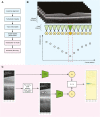
Methods: Rod intercept time (RIT) was measured in participants with and without AMD at 5 degrees from the fovea, and macular SD-OCT images were obtained. A deep learning model was trained with anatomically restricted information using a single representative B-scan through the fovea of each eye. Mean-occlusion masking was utilized to isolate the relevant imaging features.
Results: The model identified hyporeflective outer retinal bands on macular SD-OCT associated with delayed RMDA. The validation mean standard error (MSE) registered to the foveal B-scan localized the lowest error to 0.5 mm temporal to the fovea center, within an overall low-error region across the rod-free zone and adjoining parafovea. Mean absolute error (MAE) on the test set was 4.71 minutes (8.8% of the dynamic range).
Conclusions: We report a novel framework for imaging biomarker discovery using deep learning and demonstrate its ability to identify and localize a previously undescribed biomarker in retinal imaging. The hyporeflective outer retinal bands in central macula on SD-OCT demonstrate a structural basis for dysfunctional rod vision that correlates to published histopathologic findings.
Translational relevance: This agnostic approach to anatomic biomarker discovery strengthens the rationale for RMDA as an outcome measure in early AMD clinical trials, and also expands the utility of deep learning beyond automated diagnosis to fundamental discovery.
Keywords: age-related macular degeneration; biomarker; deep learning; drusen; rod-mediated dark adaptation; spectral domain optical coherence tomography.
Copyright 2020 The Authors.
Conflict of interest statement
Disclosure: A.Y. Lee, US Food and Drug Administration (E), grants from Santen (F), Carl Zeiss Meditec (F), and Novartis (F), personal fees from Genentech (R), Topcon (R), and Verana Health (R), outside of the submitted work. This article does not reflect the opinions of the Food and Drug Administration; C.S. Lee, None; M.S. Blazes, None; J.P. Owen, None; Y. Bagdasarova, None; Y. Wu, None; T. Spaide, None; R.T. Yanagihara, None; Y. Kihara, None; M.E. Clark, None; M.Y. Kwon, None; C. Owsley, is an inventor on the device used to measure dark adaptation in this study; C.A. Curcio, is a stockholder in MacRegen Inc.
Figures


References
-
- Friedman DS, O'Colmain BJ, Muñoz B, et al. .. Prevalence of age-related macular degeneration in the United States. Arch Ophthalmol. 2004; 122(4): 564–572. - PubMed
-
- Rosenfeld PJ, Brown DM, Heier JS, et al. .. Ranibizumab for neovascular age-related macular degeneration. N Engl J Med. 2006; 355(14): 1419–1431. - PubMed
-
- Brown DM, Kaiser PK, Michels M, et al. .. Ranibizumab versus verteporfin for neovascular age-related macular degeneration. N Engl J Med. 2006; 355(14): 1432–1444. - PubMed
Publication types
MeSH terms
Grants and funding
LinkOut - more resources
Full Text Sources
Medical

