. 2020 Dec 10:27:25-32.
doi: 10.1016/j.jot.2020.10.009. eCollection 2021 Mar.

De-osteogenic-differentiated mesenchymal stem cells accelerate fracture healing by mir-92b

Affiliations

De-osteogenic-differentiated mesenchymal stem cells accelerate fracture healing by mir-92b

Yonghui Hou et al. J Orthop Translat. .

Abstract

Background: Mesenchymal stem cells (MSCs) are promising targets for therapeutic use in regenerative medicine and tissue engineering. In the previous study, we have found that MSCs could be reverted to a primitive stem cell population after in vitro induction of osteogenic and de-osteogenic differentiation (de-osteogenic differentiated MSCs, De-Os-MSCs). De-Os-MSCs showed improved cell survival and osteogenic potential. However, the underlying mechanism and its potential effect on fracture healing has not been explored.

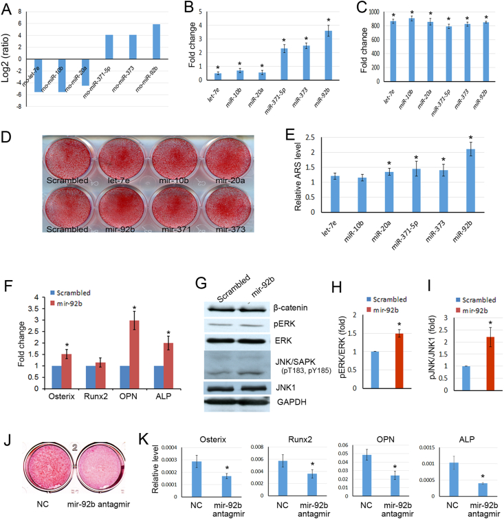
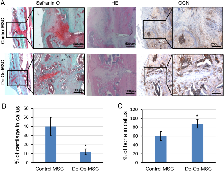
Methods: MSCs were isolated from the rat bone marrow. MicroRNAs were cloned into lentiviral vectors and transduced into MSCs to observe the effects on osteogenesis. The expression levels of marker genes were evaluated by quantitative RT-PCR. Ectopic bone formation model was used to evaluate the bone regeneration ability of mir-92b transduced MSCs in vivo. An open femur fracture model was established, and MSCs or De-Os-MSCs were administrated to the fracture sites. Histological, biomechanical and microCT analysis were used to evaluate the quality of bone.

Results: In the present study, we found that mir-92b was significantly increased in the secretions of De-Os-MSCs. And mir-92b could promote the osteogenic differentiation potential of MSCs by activating pERK and JNK signaling pathways. The ectopic bone formation assay showed that MSCs overexpressing mir-92b formed more bone like tissues in vivo. Most importantly, we found local administration of De-Os-MSCs could accelerate fracture healing using an open femur fracture model in rats. The quality of bone property was much better as shown by microCT and biomechanical testing.

Conclusion: Taken together, our study demonstrated that mir-92b promoted osteogenesis of MSCs, which was partially accounted for the enhanced osteogenic differentiation potential of De-Os-MSCs. And De-Os-MSCs had shown better regenerative capacity in accelerating fracture healing when they were locally given.

The translational potential of this article: De-Os-MSCs could be used to accelerate fracture healing, and reduce the occurrence of delayed unions and non-unions.

Keywords: De-Os-MSCs; Fracture; Mesenchymal stem cells; Mir-92b.


Conflict of interest statement

The authors have no conflicts of interest to disclose in relation to this article.

Figures

Fig. 1
Fig. 1
Mir-92b promoted osteogenesis in MSCs (A–B) The top three up/down-regulated microRNAs in De-Os-MSCs were listed, and verified by qPCR (B) (C–E) The scrambled control, let-7e, mir-10b, mir-20a, mir-92b, mir-371 and mir-373 were transduced into MSCs with lentiviruses. The overexpression of each microRNA was verified by qPCR (C). The infected MSCs were induced to undergo osteogenic differentiation for 10 days, then the calcium deposits were stained with Alizarin Red S (D), and quantified (E) (F) Total RNA was extracted from MSCs infected with mir-92b or scrambled control. The mRNA expression levels of Osterix, Runx2, OPN and ALP were detected by qPCR. β-actin was used as an internal control. The data was expressed as mean ​± ​SD (n ​= ​3). ∗p ​< ​0.05 (G–I) Total proteins were extracted from MSCs transduced with scrambled control or mir-92b. Then the proteins were analyzed by western blot using indicated antibodies. The protein levels of pERK (H) and pJNK (I) was normalized to ERK and JNK1 respectively. All the data represent mean ​± ​SD of three independent experiments. ∗p ​< ​0.05 (J–K) The mir-92b antagmir was transfected into MSCs, then the cells were treated with osteogenic induction medium for 10 days, the calcium deposits were stained with Alizarin Red S (J), the changes of osteogenesis-related genes was checked by qPCR (K).
Fig. 2
Fig. 2
Overexpression of mir-92b in MSCs promoted ectopic bone formation. MSCs transduced with scrambled/GFP or mir-92b/GFP were loaded onto sterilized porous calcium phosphate restorable granules, and then implanted subcutaneously into the dorsal surfaces. The transplants were harvested 8 weeks later for histological examination (A) The sections were stained with routine hematoxylin and eosin, amorphous osteoid matrix could be seen (arrow head). At: adipose tiusse; ft: fibrous tissue. Scale bar ​= ​400 ​μm (B) Quantification of osteoid matrix showed that there was more osteoid matrix in mir-92b overexpressing group. Five microscopic fields from each sample were used for measurement. Results are presented as mean ​± ​SD, ∗p ​< ​0.05.
Fig. 3
Fig. 3
De-Os-MSCs increased the cartilaginous content in callus at 3 weeks after fracture (A) At 3 weeks after fracture, the femurs were collected for histological analysis. Longitudinal sections of callus were subjected to HE, Safranin O staining and immunohistochemical analysis using GFP antibody (B) The relative cartilaginous area in the callus was measured according to the Safranin O staining (C) The number of locally injected MSCs in callus was counted (n ​= ​3). The data represented mean ​± ​SD. ∗p ​< ​0.05.
Fig. 4
Fig. 4
Micro-CT analysis of fracture callus. At 8 weeks after fracture, the femurs were collected for micro-CT analysis (A) Representative 3D images generated from micro-CT analysis of femur fracture healing in rats transplanted with De-Os-MSCs and normal MSCs (B–D) The bone volume density-BVt/TV and BVl/TV was analyzed by micro-CT. TV: Total tissue volume; BVt: Total bone volume; BVh: Volume of high-density bone; BVl: volume of low-density bone. ∗p ​< ​0.05, n ​= ​4.
Fig. 5
Fig. 5
Histological staining of fracture callus. At 8 weeks after fracture, the femurs were collected for histological analysis (A) Representative images of Safranin O, HE staining and immunohistological staining of OCN in femur fracture callus of rats transplanted with De-Os-MSCs and normal MSCs (B&C) The relative cartilaginous area and newly formed bone in the callus was measured according to the staining. The data represented mean ​± ​SD. ∗p ​< ​0.05.
Fig. 6
Fig. 6
Mechanical test analysis of fracture callus. At 8 weeks after fracture, the femurs were collected for mechanical test analysis (A–C) Mechanical properties of the fractured bones by four-point bending test. The mechanical properties (including ultimate load, energy to failure and stiffness) in the fractured femur were normalized with contra-lateral intact femur (in percent). The data represented mean ​± ​SD. ∗p ​< ​0.05, n ​= ​5.
Supplementary Fig. 1
The pre-mature mir-92b was cloned into a lentiviral vector labeled with GFP, and packaged into pseudo-lentivirus. Then the MSCs were transduced with lentivirus to overexpress mir-92b. The expression of GFP was viewed under fluorescent microscope.

References

    1. Einhorn T.A., Gerstenfeld L.C. Fracture healing: mechanisms and interventions. Nat Rev Rheumatol. 2014;11(1):45–54. - PMC - PubMed
    1. Marsell R., Einhorn T.A. The biology of fracture healing. Injury. 2011;42(6):551–555. - PMC - PubMed
    1. Axelrad T.W., Kakar S., Einhorn T.A. New technologies for the enhancement of skeletal repair. Injury. 2007;38(Suppl 1):S49–S62. - PubMed
    1. Kon E., Filardo G., Roffi A., Di Martino A., Hamdan M., De Pasqual L. Bone regeneration with mesenchymal stem cells. Clin Cases Miner Bone Metab. 2012;9(1):24–27. - PMC - PubMed
    1. Parekkadan B., Milwid J.M. Mesenchymal stem cells as therapeutics. Annu Rev Biomed Eng. 2010;12:87–117. - PMC - PubMed

LinkOut - more resources