. 2020 Nov 8;11(6):948-952.
doi: 10.4103/idoj.IDOJ_100_20. eCollection 2020 Nov-Dec.

Lupus Vulgaris in Darker Skin: Dermoscopic and Histopathologic Incongruity

Affiliations

Lupus Vulgaris in Darker Skin: Dermoscopic and Histopathologic Incongruity

Balachandra S Ankad et al. Indian Dermatol Online J. .

Abstract

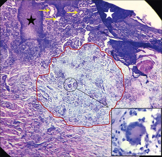
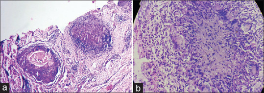
Introduction: Lupus Vulgaris (LV) is the chronic, progressive, tissue destructive form of cutaneous tuberculosis. LV should be diagnosed and treated to prevent scaring and deformities. Histopathology is the gold standard for the diagnosis. Dermoscopy is helpful tool in diagnosing different dermatological condition. Here, dermoscopic and histopathogical correlation in LV was attempted.

Materials and methods: It was a cross sectional, observational study done from February 2019 to October 2019. Nineteen patients of LV were included. Dermlite 4 with attached smart phone (iphone) was employed. LV lesions were subjected to skin biopsy to confirm the diagnosis.

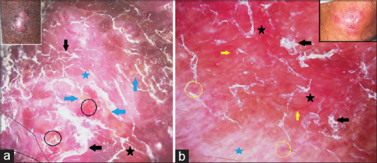
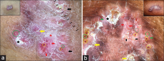
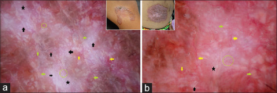
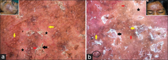
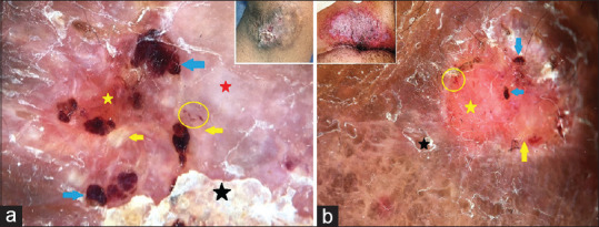
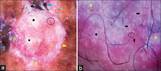
Results: Study enrolled 19 patients, with 8males, 5 female and 6 children. Dermoscopy showed yellowish-white globules, white structureless areas and white scales were noted in 19 (100%) patients. Telangiectasias were seen in 16 (84.21%) patients as long linear, branching and short linear vessels. Pinkish-red background was noted in all patients (100% n=19). Newer observations included white shiny streaks, white rosettes and bluish hue. Age, sex, duration of lesions had no influence in the dermoscopic patterns. Discrepancy in dermoscopic-histopathologic correlation was noted. Facial lesions showed increased frequency of follicular plugs, patulous follicles and white rosettes.

Conclusion: Dermoscopy is widely gaining importance in the realm of dermatology. In this study, dermoscopy demonstrated characteristic patterns in LV. Thus, dermoscopy a non-invasive procedure can be used as diagnostic tool in many infective dermatoses.

Keywords: Dermoscopy; diagnosis; histopathology; lupus vulgaris; patterns.


Conflict of interest statement

There are no conflicts of interest.

Figures

Figure 1
Figure 1
Panels a and b: Dermoscopy of lupus vulgaris shows yellow globules (blue arrows), white scales (black arrows), white structureless areas (blue stars), dotted vessels (black circles) and red globules (yellow circles) on the pinkish background (black stars). Linear vessels (yellow arrows) are well appreciated. Inset: Clinical images. [Polarized Dermoscopy, original magnification ×10]
Figure 2
Figure 2
Panels a and b: Dermoscopy of lupus vulgaris shows yellow globules (black arrows), white scales (black stars), white structureless areas (red star), red dots (green circles), red globules (yellow circles) and linear vessels (green arrows). Note the white shiny streaks (red arrows) and pinkish background. Follicular plugs (yellow arrows) are well appreciated. Inset: Clinical images. [Polarized dermoscopy, original magnification ×10]
Figure 3
Figure 3
Panels a and b: Dermoscopy of lupus vulgaris shows white shiny streaks (black arrows), white structureless areas (black stars), linear and branching vessels (yellow arrows) and dotted vessels (green circles). Yellow globules (green arrows) and pinkish background are well appreciated. Inset: Clinical images. [Polarized dermoscopy, original magnification ×10]
Figure 4
Figure 4
Panels a and b: Dermoscopy of lupus vulgaris shows white scales (black arrows), white structureless areas (black stars), linear vessels (yellow arrows) and patulous follicular openings (red arrows) on pinkish background. Note the four-dot clod (black circle) in panel b. Inset: Clinical images. [Polarized dermoscopy, original magnification ×10]
Figure 5
Figure 5
Panels a and b: Dermoscopy of lupus vulgaris shows yellow globules (yellow arrows), ulceration (yellow stars), white structureless areas (red star), red globules (blue arrows) and red dots (yellow circles). White scales (black stars) and pinkish background are well appreciated. Inset: Clinical images. [Polarized dermoscopy, original magnification ×10]
Figure 6
Figure 6
Panels a and b: Dermoscopy of lupus vulgaris shows white structureless areas (black stars), ulceration (yellow arrows), dark red globules (blue arrows), red dots and globules (black circles). Note the bluish hue (yellow stars), pinkish background and white rosette (red arrow) and white scales (green arrow). [Polarized dermoscopy, original magnification ×10]
Figure 7
Figure 7
Histopathology of lupus vulgaris shows acanthosis (black star), hyperkeratosis (white star) and dilated capillaries (yellow arrows) and granuloma (red circle). Inset: Langhans giant cell. [H and E, 10×]
Figure 8
Figure 8
Panels a and b: Histopathology of lupus vulgaris shows well-demarcated granuloma with dilated capillaries and dilated follicles with follicular plug and lymphocytic infiltrate (panel a). Granuloma with multiple Langhans giant cells and lymphocytes are well appreciated (panel b). [H and E, 10×]

References

    1. Pai VV, Naveen KN, Athanikar SB, Dinesh US, Divyashree A, Gupta G. A clinico-histological study of Lupus vulgaris: A 3 year experience at a tertiary care centre. Indian Dermatol Online J. 2014;5:461–5. - PMC - PubMed
    1. Kittler H, Pehamberger H, Wolff K, Binder M. Diagnostic accuracy of dermoscopy. Lancet Oncol. 2002;3:159–65. - PubMed
    1. Errichetti E, Stinco G. Dermoscopy in general dermatology: A practical overview. Dermatol Ther (Heidelb) 2016;6:471–507. - PMC - PubMed
    1. Brasiello M, Zalaudek I, Ferrara G, Gourhant JY, Capoluongo P, Roma P, et al. Lupus vulgaris: A new look at an old symptom-The lupoma observed with dermoscopy. Dermatology. 2009;218:172–4. - PubMed
    1. Kreusch J. How to perform dermoscopy of non-pigmented skin lesions. In: Zalaudek I, Argenziano G, Giacomel J, editors. Dermatoscopy of Non-Pigmented Skin Tumors. London: CRC Press; 2016. pp. 17–8.