Review
. 2020 Dec 3:7:570689.
doi: 10.3389/fcvm.2020.570689. eCollection 2020.

Multimodality Imaging for Discordant Low-Gradient Aortic Stenosis: Assessing the Valve and the Myocardium

Affiliations
Review

Multimodality Imaging for Discordant Low-Gradient Aortic Stenosis: Assessing the Valve and the Myocardium

Ezequiel Guzzetti et al. Front Cardiovasc Med. .

Abstract

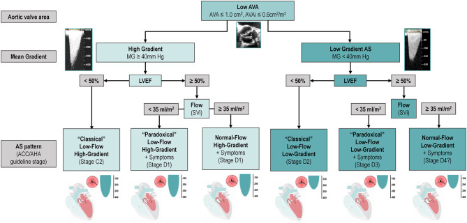
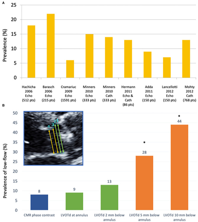
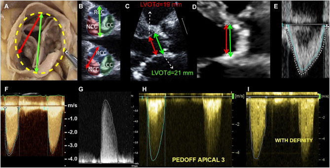
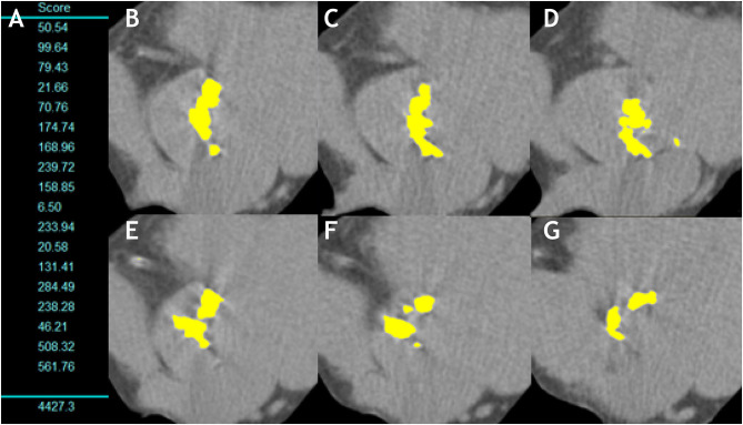
Aortic stenosis (AS) is a disease of the valve and the myocardium. A correct assessment of the valve disease severity is key to define the need for aortic valve replacement (AVR), but a better understanding of the myocardial consequences of the increased afterload is paramount to optimize the timing of the intervention. Transthoracic echocardiography remains the cornerstone of AS assessment, as it is universally available, and it allows a comprehensive structural and hemodynamic evaluation of both the aortic valve and the rest of the heart. However, it may not be sufficient as a significant proportion of patients with severe AS presents with discordant grading (i.e., an AVA ≤ 1 cm2 and a mean gradient <40 mmHg) which raises uncertainty about the true severity of AS and the need for AVR. Several imaging modalities (transesophageal or stress echocardiography, computed tomography, cardiovascular magnetic resonance, positron emission tomography) exist that allow a detailed assessment of the stenotic aortic valve and the myocardial remodeling response. This review aims to provide an updated overview of these multimodality imaging techniques and seeks to highlight a practical approach to help clinical decision making in the challenging group of patients with discordant low-gradient AS.

Keywords: aortic stenosis; computed tomography; echocardiography; low-gradient aortic stenosis; magnetic resonance imaging.


Conflict of interest statement

The authors declare that the research was conducted in the absence of any commercial or financial relationships that could be construed as a potential conflict of interest.

Figures

Figure 1
Figure 1
Classification and Characterization of the different types of AS according to AVA, Gradient, LVEF and Flow. The classification of types of AS, including only the categories associated with symptoms and/or depressed LVEF. It does not include stage C1 (i.e., patients with high-gradient AS, no symptoms, and preserved LVEF). Question mark indicates stage labels that are proposed by the authors but are not included in the guidelines and will need to be further tested and validated. ACC, American College of Cardiology; AHA, American Heart Association; AS, aortic stenosis; AVA, aortic valve area; AVAi, indexed aortic valve area; LVEF, left ventricular ejection fraction; MG, mean gradient; SVi, stroke volume index.
Figure 2
Figure 2
Prevalence of low flow according to different studies and measurement techniques. (A) Prevalence of paradoxical low-flow severe AS (i.e., AVA <1.0 cm2; MG <40 mmHg, LVEF>50%; SVi <35 mL/m2) according to different studies using echocardiography (Echo) and/or invasive catheterization (Cath). (B) Prevalence of low-flow state (stroke volume index ≤ 35 ml/m2) according to different measurement sites of the LVOT: (1) at the hinge points of the aortic valve leaflets (annular level); (2) very close to (i.e., 2 mm below) the annular level; (3) 5 mm below the annular level; and (4) 10 mm below the annular level, as compared to the referent standard (phase-contrast CMR) [modified from JASE (16)]. CMR, Cardiovascular magnetic resonance; LVOTd, left ventricular outflow tract diameter. *p < 0.01 as compared to CMR-PC (referent method).
Figure 3
Figure 3
Echocardiographic measurement pitfalls and how to avoid them. LVOT diameter must be measured at its maximal dimension, which generally corresponds to the bisection between the right coronary cusp hinge point anteriorly and the interleaflet triangle between the left and non-coronary cusps posteriorly (A,B). LVOTd must be measured at the annulus and not 5–10 mm below, as this leads to significant underestimation of AVA and SV (C). In this case, measuring 5 mm below the annulus (as recommended in guidelines) lead to an LVOT area of 2.83 cm2, as compared to an area of 3.46 cm2 when measured at the annulus (18% underestimation, therefore leading to significant underestimation of SV and AVA). In case of LVOT ectopic calcification, if the plane that bisects the largest diameter cannot exclude the calcium, LVOT diameter measurement should include the calcium in the measurement (D). LVOT velocity-time integral should be measured at the modal velocity (the densest line of the Pulse Wave Doppler) since flow at the LVOT is laminar (E: blue trace represents the modal velocity whereas the dashed white line overestimates LVOT VTI). For an accurate measurement of the transaortic jet velocity, tracing should be done at peak velocities but excluding fine linear signals (F: green trace represents the correct measurement, whereas the dashed white line overestimates aortic valve VTI by including linear signals). It is paramount that the Doppler beam is optimally aligned parallel to the stenotic aortic jet. Therefore, a meticulous search of the highest transvalvular velocity is mandatory. This requires a comprehensive Doppler study that is not only limited to the apical window but also includes right parasternal, suprasternal, and sometimes subcostal approaches using a small, dedicated CW Doppler transducer (pencil probe or Pedoff transducer) (G,H). Finally, the use of ultrasound enhancing agents (i.e., contrast echocardiography as with Definity®) might lead to overestimation of transvalvular velocities and gradients and therefore caution should be taken (I).
Figure 4
Figure 4
Aortic valve calcification measurement in a man with severe aortic stenosis. (A) Shows the measurement of the aortic valve calcium in each 3-mm slice and total sum (AVC score = 4,427.3AU). (B–G) Show the multiple axial images from aortic annulus to aortic root with any aortic valve calcification highlighted in yellow by the software.
Figure 5
Figure 5
Proposed diagnostic algorithm for severe aortic stenosis. Proposed algorithm for diagnostic assessment of AS. As compared to ESC/EACTS guideline algorithm (7), we suggest assessing SVi (and/or Q mean) in all AS patients (regardless of mean gradient). After careful revision of accuracy of echocardiographic measurements (left box), MDCT AVC is proposed to confirm true anatomical severity in all discordant grading patients (i.e., low AVA and low gradient/Vmax) regardless of LVEF and SVi. Dobutamine stress echocardiography remains as an alternative option if MDCT unavailable (see text). AS, aortic stenosis; AVA, aortic valve area; AVC, aortic valve calcium score; DVI, Doppler velocity index; MDCT, multidetector computed tomography; MG, mean transvalvular gradient; SV, stroke volume, LVOT, left ventricular outflow tract, Q mean, mean transvalvular flow rate, Vmax, peak aortic velocity.
Figure 6
Figure 6
Multimodality imaging assessment of discordant low-gradient aortic stenosis. In (A), a 75 year-old man with the classical form of low-flow, low-gradient AS i.e. with a left ventricular ejection fraction of 30%. The patient had an aortic valve area (AVA) <1.0, in discordance with a mean pressure gradient (MG) <40 mmHg (B). Usually, dobutamine stress echocardiography allows to assess MG/AVA at flow normalizaion. However, as shown in (C), and as happens in 30–40% of the patients, the discordance persisted, which was due to a minimal increase in transvalvular flow (Qmean). In these cases, it is recommended to measure the aortic valve calcification following the Agatston method and using sex-specific cutpoints (1200 AU for women and 2000 AU for men). This eventually allowed to confirm stenosis severity (D). In (E), a woman with mild symptoms (NYHA I-II) and discordant a priori severe AS but normal LVEF and normal stroke volume (F). The AVC score is the primary approach in this subset of patients, as illustrated in the present case (G and confirmation of stenosis severity). Patients with normal LVEF are at lower risk than CLF patients. However, risk-stratification can be achieved using gadolinium enhanced cardiac magnetic resonance or NTproBNP. This patient exhibited focal myocardial fibrosis on CMR (H). Also, her NT-proBNP was measured at 660 pg/ml i.e., 7-fold the upper reference level for age and sex. Both results indicate a high-risk profile and suggest that aortic valve replacement is a reasonable option.
Figure 7
Figure 7
Cardiac damage staging system. The proposed cardiac damage staging scheme originally proposed by Généreux et al. (6) and modified by Tastet et al. (5) is based on a multi-parameter approach using echocardiographic parameters. The patient qualifies for a given stage if at least one of the proposed criteria for this stage is met. *Parameters added or modified by Tastet et al. to the original Génereux scheme for staging in asymptomatic patients with moderate or severe AS.

Similar articles

Cited by

References

    1. Clavel MA, Burwash IG, Pibarot P. Cardiac imaging for assessing low-gradient severe aortic stenosis. JACC Cardiovasc Imaging. (2017) 10:185–202. 10.1016/j.jcmg.2017.01.002 - DOI - PubMed
    1. Berthelot-Richer M, Pibarot P, Capoulade R, Dumesnil JG, Dahou A, Thébault C, et al. . Discordant grading of aortic stenosis severity: echocardiographic predictors of survival benefit associated with aortic valve replacement. JACC Cardiovasc Imaging. (2016) 9:797–805. 10.1016/j.jcmg.2015.09.026 - DOI - PubMed
    1. Dweck MR, Boon NA, Newby DE. Calcific aortic stenosis: a disease of the valve and the myocardium. J Am Coll Cardiol. (2012) 60:1854–63. 10.1016/j.jacc.2012.02.093 - DOI - PubMed
    1. Treibel TA, Badiani S, Lloyd G, Moon JC. Multimodality imaging markers of adverse myocardial remodeling in aortic stenosis. JACC Cardiovasc Imaging. (2019) 12:1532–48. 10.1016/j.jcmg.2019.02.034 - DOI - PubMed
    1. Tastet L, Tribouilloy C, Maréchaux S, Vollema EM, Delgado V, Salaun E, et al. . Staging cardiac damage in patients with asymptomatic aortic valve stenosis. J Am Coll Cardiol. (2019) 74:550–63. 10.1016/j.jacc.2019.04.065 - DOI - PubMed