. 2020 Dec 2:7:609443.
doi: 10.3389/fvets.2020.609443. eCollection 2020.

The Suppression of Th1 Response by Inducing TGF-β1 From Regulatory T Cells in Bovine Mycoplasmosis

Affiliations

The Suppression of Th1 Response by Inducing TGF-β1 From Regulatory T Cells in Bovine Mycoplasmosis

Yamato Sajiki et al. Front Vet Sci. .

Abstract

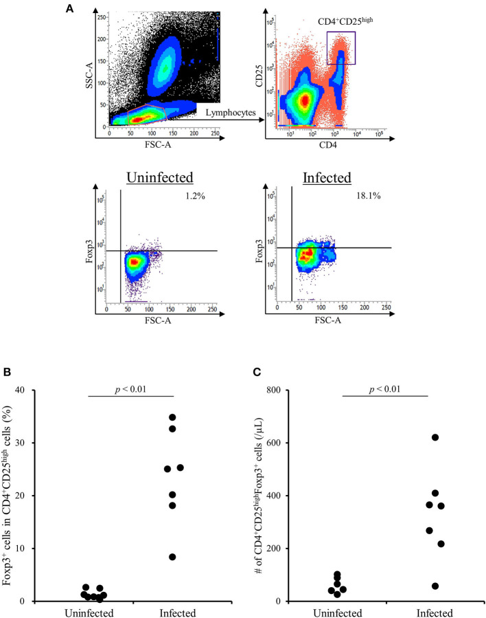
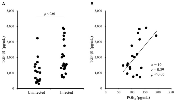
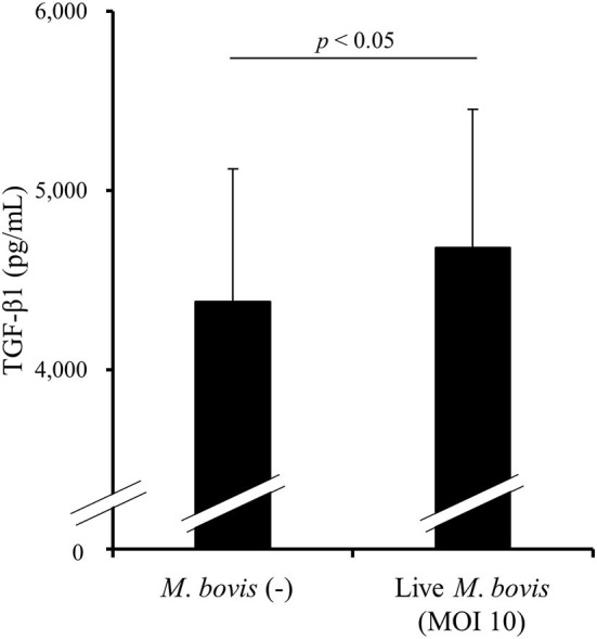
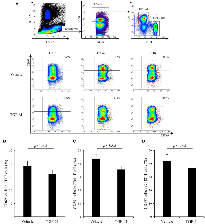
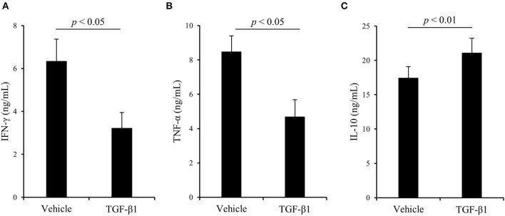
Regulatory T cells (Tregs) regulate immune responses and maintain host immune homeostasis. Tregs contribute to the disease progression of several chronic infections by oversuppressing immune responses via the secretion of immunosuppressive cytokines, such as transforming growth factor (TGF)-β and interleukin-10. In the present study, we examined the association of Tregs with Mycoplasma bovis infection, in which immunosuppression is frequently observed. Compared with uninfected cattle, the percentage of Tregs, CD4+CD25highFoxp3+ T cells, was increased in M. bovis-infected cattle. Additionally, the plasma of M. bovis-infected cattle contained the high concentrations of TGF-β1, and M. bovis infection induced TGF-β1 production from bovine immune cells in in vitro cultures. Finally, we analyzed the immunosuppressive effects of TGF-β1 on bovine immune cells. Treatment with TGF-β1 significantly decreased the expression of CD69, an activation marker, in T cells, and Th1 cytokine production in vitro. These results suggest that the increase in Tregs and TGF-β1 secretion could be one of the immunosuppressive mechanisms and that lead to increased susceptibility to other infections in terms of exacerbation of disease during M. bovis infection.

Keywords: Mycoplasma bovis; TGF-β1; cattle; immunosuppression; regulatory T cell.


Conflict of interest statement

The authors declare that the research was conducted in the absence of any commercial or financial relationships that could be construed as a potential conflict of interest.

Figures

Figure 1
Figure 1
The increase of CD4+CD25highFoxp3+ T cells in M. bovis-infected cattle. (A) The gating strategy and representative plots for Treg staining. (B) The percentage of Foxp3+ cells in CD4+CD25high T cells in M. bovis-infected (n = 7) and -uninfected (n = 8) cattle. (C) The number of CD4+CD25highFoxp3+ cells in M. bovis-infected (n = 7) and -uninfected (n = 6) cattle. (B,C) Statistical significance was determined by the Mann-Whitney U test. Uninfected, M. bovis-uninfected cattle; Infected, M. bovis-infected cattle.
Figure 2
Figure 2
The increase of TGF-β1 concentrations in blood plasma of M. bovis-infected cattle. (A) TGF-β1 concentrations in the plasma from M. bovis-infected (n = 22) and -uninfected (n = 16) cattle were determined by ELISA. Statistical significance was determined by the Mann-Whitney U test. (B) The correlation between TGF-β1 and PGE2 concentrations in the plasma of M. bovis-infected cattle (n = 19). Correlation statistic was analyzed using the Spearman correlation.
Figure 3
Figure 3
The induction of TGF-β1 production by M. bovis. PBMCs from uninfected cattle were cultured with live M. bovis, and TGF-β1 concentrations in culture supernatants were measured by ELISA (n = 8). Data are presented as means, and the error bars indicate standard errors. Statistical significance was determined by the Wilcoxon signed-rank test. MOI, multiplicity of infection.
Figure 4
Figure 4
The effect of TGF-β1 on CD69 expression in cattle. (A–D) PBMCs from uninfected cattle (n = 7) were incubated with TGF-β1 in the presence of Con A. (A) The gating strategy and representative plots for CD69 expression. (B–D) CD69 expression in T cells (B), CD4+ T cells (C), and CD8+ T cells (D) were measured by flow cytometry. Data are presented as means, and the error bars indicate standard errors. Statistical significance was determined by the Wilcoxon signed-rank test.
Figure 5
Figure 5
The effect of TGF-β1 on cytokine production in cattle. (A–C) PBMCs from uninfected cattle (n = 7) were incubated with TGF-β1 in the presence of Con A. After incubation, IFN-γ (A), TNF-α (B), and IL-10 (C) concentrations in culture supernatants were determined by ELISA. Data are presented as means, and the error bars indicate standard errors. Statistical significance was determined by the Wilcoxon signed-rank test.

Similar articles

Cited by

References

    1. Janardhan KS, Hays M, Dyer N, Oberst RD, Debey BM. Mycoplasma bovis outbreak in a herd of North American bison (Bison bison). J Vet Diagn Invest. (2010) 22:797–801. 10.1177/104063871002200528 - DOI - PubMed
    1. Nicholas RA. Bovine mycoplasmosis: silent and deadly. Vet Rec. (2011) 168:459–62. 10.1136/vr.d2468 - DOI - PubMed
    1. Higuchi H, Iwano H, Gondaira S, Kawai K, Nagahata H. Prevalence of Mycoplasma species in bulk tank milk in Japan. Vet Rec. (2011) 169:442. 10.1136/vr.d5331 - DOI - PubMed
    1. Higuchi H, Gondaira S, Iwano H, Hirose K, Nakajima K, Kawai K, et al. . Mycoplasma species isolated from intramammary infection of Japanese dairy cows. Vet Rec. (2013) 172:557. 10.1136/vr.101228 - DOI - PubMed
    1. Gagea MI, Bateman KG, Shanahan RA, van Dreumel T, McEwen BJ, Carman S, et al. . Naturally occurring Mycoplasma bovis-associated pneumonia and polyarthritis in feedlot beef calves. J Vet Diagn Invest. (2006) 18:29–40. 10.1177/104063870601800105 - DOI - PubMed