

Large leiomyoma of lower esophagus diagnosed by endoscopic ultrasonography-fine needle aspiration: A case report
- PMID: 33344578
- PMCID: PMC7716314
- DOI: 10.12998/wjcc.v8.i22.5809
Large leiomyoma of lower esophagus diagnosed by endoscopic ultrasonography-fine needle aspiration: A case report
Abstract
Background: Benign esophageal tumors are rare accounting for < 1% of esophageal tumors; two-thirds of which are leiomyomas. Esophageal leiomyoma is a benign tumor derived from mesenchymal tissue that is completely muscularly differentiated. Most esophageal leiomyomas are < 5 cm. Esophageal leiomyomas > 5 cm are rare. We describe a case of a large esophageal leiomyoma involving the cardia and diaphragm.
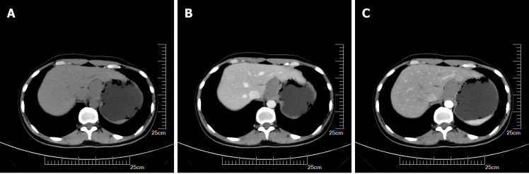
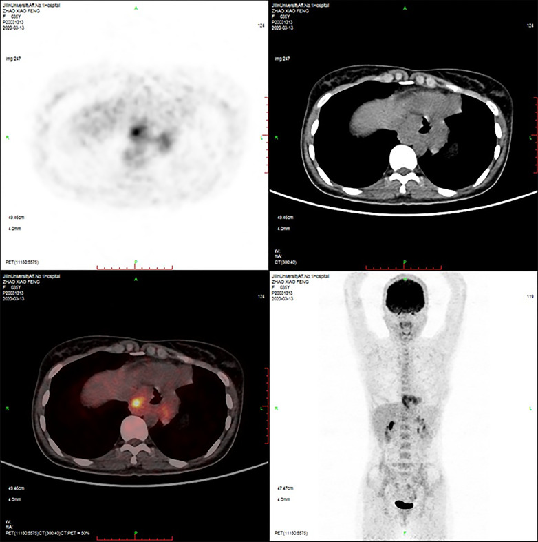
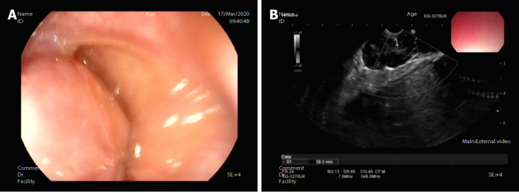
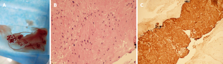
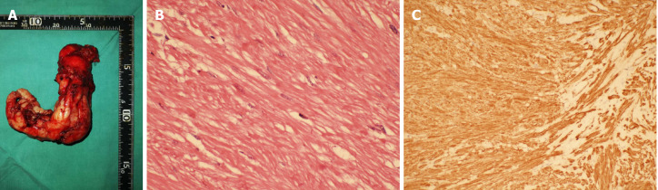
Case summary: A 35-year-old woman presented to the doctor because of a choking sensation after eating. Physical examination showed no positive signs. Gastroscopy indicated an uplifted change in the cardia. Enhanced computed tomography revealed space-occupying lesions in the lower part of the esophagus and cardia, which were likely to be malignant. Positron emission tomography-computed tomography showed increased metabolism of soft tissue masses in the lower esophagus and near the cardia. Malignant lesions were considered, and mesenchymal tumors were not excluded. Endoscopic ultrasonography was performed to examine a hypoechoic mass in the lower esophagus, which was unclear from the esophageal wall. Clinical evaluation suggested diagnosis of esophageal and cardiac stromal tumors. Finally, histological specimens obtained by endoscopic ultrasonography- fine needle aspiration suggested leiomyoma. The patient underwent laparoscopic local resection of the tumor. The postoperative pathological diagnosis was leiomyoma.
Conclusion: Endoscopic ultrasonography-fine needle aspiration is necessary for the diagnosis of gastrointestinal leiomyomas. It provides a strong basis for diagnosis of gastrointestinal tumors of unknown nature and origin.
Keywords: Case report; Endoscopic ultrasonography; Endoscopic ultrasonography-fine needle aspiration; Esophageal tumor; Fine needle aspiration; Leiomyoma.
©The Author(s) 2020. Published by Baishideng Publishing Group Inc. All rights reserved.
Conflict of interest statement
Conflict-of-interest statement: There are no conflicts of interest by any other the authors of this manuscript including Drs. Min Rao, Qing-Qing Meng, and Pu-Jun Gao.
Figures
Similar articles
-
Are biopsies during endoscopic ultrasonography necessary for a suspected esophageal leiomyoma? Is laparoscopy always feasible?World J Clin Cases. 2023 Mar 26;11(9):2116-2118. doi: 10.12998/wjcc.v11.i9.2116. World J Clin Cases. 2023. PMID: 36998946 Free PMC article.
-
Endoscopic Ultrasonography in the Diagnosis and Treatment Strategy Choice of Esophageal Leiomyoma.Clinics (Sao Paulo). 2017 Apr;72(4):197-201. doi: 10.6061/clinics/2017(04)01. Clinics (Sao Paulo). 2017. PMID: 28492717 Free PMC article.
-
Successful en bloc endoscopic full-thickness resection of a giant cervical esophageal leiomyoma originating from muscularis propria.J Cardiothorac Surg. 2019 Jan 21;14(1):16. doi: 10.1186/s13019-019-0847-5. J Cardiothorac Surg. 2019. PMID: 30665433 Free PMC article.
-
Difficult endoscopic resection of a giant esophageal fibrovascular polyp: case report and literature review.J Int Med Res. 2021 Aug;49(8):3000605211039801. doi: 10.1177/03000605211039801. J Int Med Res. 2021. PMID: 34459277 Free PMC article. Review.
-
Esophageal leiomyoma and simultaneous overlying squamous cell carcinoma: a case report and review of the literature.BMC Surg. 2021 Apr 29;21(1):221. doi: 10.1186/s12893-021-01214-2. BMC Surg. 2021. PMID: 33926432 Free PMC article. Review.
Cited by
-
Missed Giant Lower Esophageal Leiomyoma in a Young Female Presenting with Refractory Gastroesophageal Reflux Disease.Case Rep Med. 2021 Jul 20;2021:9925224. doi: 10.1155/2021/9925224. eCollection 2021. Case Rep Med. 2021. PMID: 34335788 Free PMC article.
-
Are biopsies during endoscopic ultrasonography necessary for a suspected esophageal leiomyoma? Is laparoscopy always feasible?World J Clin Cases. 2023 Mar 26;11(9):2116-2118. doi: 10.12998/wjcc.v11.i9.2116. World J Clin Cases. 2023. PMID: 36998946 Free PMC article.
-
Slicing Through the Options: A Systematic Review of Esophageal Leiomyoma Management.Cureus. 2025 Apr 2;17(4):e81614. doi: 10.7759/cureus.81614. eCollection 2025 Apr. Cureus. 2025. PMID: 40177232 Free PMC article. Review.
References
-
- Jiang W, Rice TW, Goldblum JR. Esophageal leiomyoma: experience from a single institution. Dis Esophagus. 2013;26:167–174. - PubMed
-
- De Giacomo T, Bruschini P, Arcieri S, Ruberto F, Venuta F, Diso D, Francioni F. Partial oesophagectomy for giant leiomyoma of the oesophagus: report of 7 cases. Eur J Cardiothorac Surg. 2015;47:143–145. - PubMed
-
- Shin S, Choi YS, Shim YM, Kim HK, Kim K, Kim J. Enucleation of esophageal submucosal tumors: a single institution's experience. Ann Thorac Surg. 2014;97:454–459. - PubMed
Publication types
LinkOut - more resources
Full Text Sources
