

Usefulness of ultrasonography to assess the response to steroidal therapy for the rare case of type 2b immunoglobulin G4-related sclerosing cholangitis without pancreatitis: A case report
- PMID: 33344580
- PMCID: PMC7716308
- DOI: 10.12998/wjcc.v8.i22.5821
Usefulness of ultrasonography to assess the response to steroidal therapy for the rare case of type 2b immunoglobulin G4-related sclerosing cholangitis without pancreatitis: A case report
Abstract
Background: A type 2b immunoglobulin G4 (IgG4)-related sclerosing cholangitis (SC) without autoimmune pancreatitis is a rare condition with IgG4-SC. While the variety of the imaging modalities have tested its usefulness in diagnosing the IgG4-SC, however, the usage of ultrasonography for the assessment of the response to steroidal therapy on the changes of bile duct wall thickness have not been reported in the condition. Therefore, the information of our recent case and reported cases have been summarized.
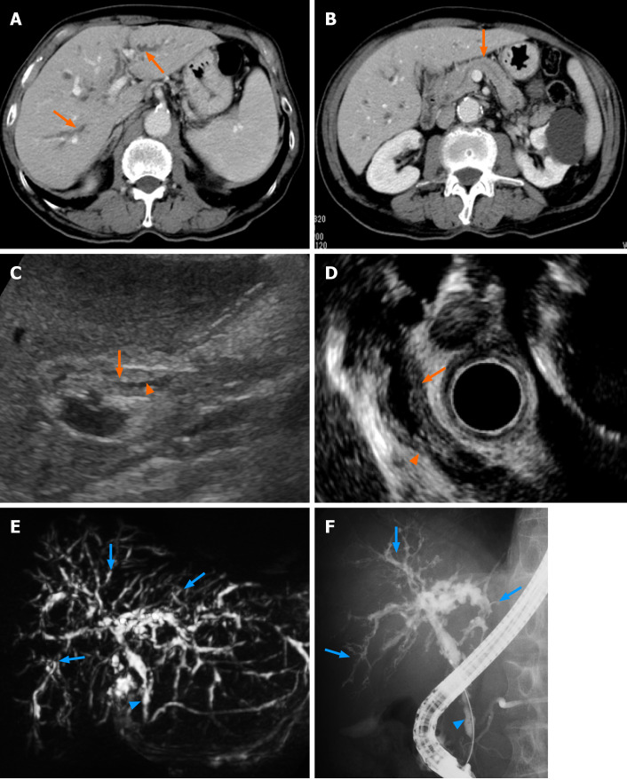
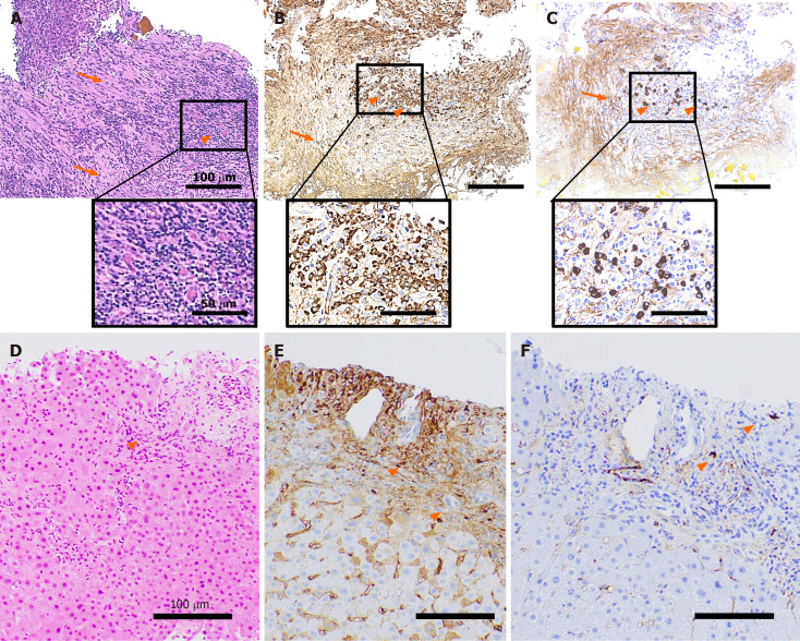
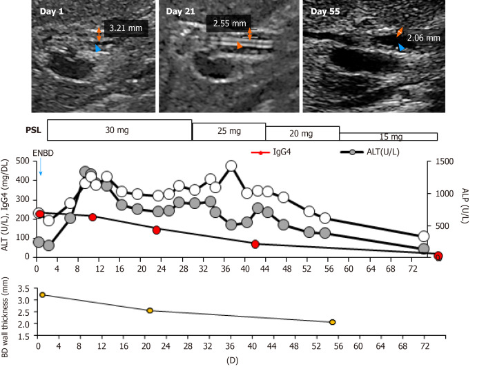
Case summary: We report the case of an 82-year-old Japanese man diagnosed with isolated IgG4-related SC based on the increase of serum IgG4, narrowing of the bile duct, its wall thickness, no complication of autoimmune pancreatitis, and IgG4 positive inflammatory cell infiltration to the wall with the fibrotic changes. The cholangiogram revealed type 2b according to the classification. Corticosteroid treatment showed a favorable effect, with the smooth decrease in serum IgG4 and the improvement of the bile duct wall thickness.
Conclusion: As isolated type 2b, IgG4-SC is rare, the images, histological findings, and clinical course of our case will be helpful for physicians to diagnose and treat the new cases appropriately.
Keywords: Autoimmune pancreatitis; Case report; Corticosteroid; Imaging; Immunoglobulin G4-related sclerosing cholangitis; Type 2b; Ultrasonography.
©The Author(s) 2020. Published by Baishideng Publishing Group Inc. All rights reserved.
Conflict of interest statement
Conflict-of-interest statement: The authors declare that they have no current financial arrangement or affiliation with any organization that may have a direct influence on their work.
Figures
Similar articles
-
Usefulness of laparoscopy and intraductal ultrasonography in a patient with isolated immunoglobulin G4-related sclerosing cholangitis.Clin J Gastroenterol. 2018 Feb;11(1):62-68. doi: 10.1007/s12328-017-0787-3. Epub 2017 Nov 1. Clin J Gastroenterol. 2018. PMID: 29094322
-
Isolated intrapancreatic IgG4-related sclerosing cholangitis.World J Gastroenterol. 2015 Jan 28;21(4):1334-43. doi: 10.3748/wjg.v21.i4.1334. World J Gastroenterol. 2015. PMID: 25632210 Free PMC article.
-
[Clinical observation of isolated immunoglobulin G4-related sclerosing cholangitis and immunoglobulin G4 sclerosing cholangitis combined autoimmune pancreatitis].Zhonghua Gan Zang Bing Za Zhi. 2018 Jun 20;26(6):415-419. doi: 10.3760/cma.j.issn.1007-3418.2018.06.005. Zhonghua Gan Zang Bing Za Zhi. 2018. PMID: 30317753 Chinese.
-
Immunoglobulin G4-related sclerosing cholangitis.J Dig Dis. 2019 Jul;20(7):357-362. doi: 10.1111/1751-2980.12789. Epub 2019 Jul 2. J Dig Dis. 2019. PMID: 31112324 Review.
-
Role of endoscopy in the diagnosis of autoimmune pancreatitis and immunoglobulin G4-related sclerosing cholangitis.Dig Endosc. 2014 Sep;26(5):627-35. doi: 10.1111/den.12289. Epub 2014 Apr 8. Dig Endosc. 2014. PMID: 24712522 Review.
Cited by
-
Current and innovative methods for the diagnosis of COVID‑19 infection (Review).Int J Mol Med. 2021 Jun;47(6):100. doi: 10.3892/ijmm.2021.4933. Epub 2021 Apr 13. Int J Mol Med. 2021. PMID: 33846767 Free PMC article. Review.
-
Secondary sclerosing cholangitis and IgG4-sclerosing cholangitis - A review of cholangiographic and ultrasound imaging.Endosc Ultrasound. 2023 Mar-Apr;12(2):181-199. doi: 10.4103/EUS-D-22-00208. Endosc Ultrasound. 2023. PMID: 36588352 Free PMC article. Review.
References
-
- Nakazawa T, Naitoh I, Hayashi K, Okumura F, Miyabe K, Yoshida M, Yamashita H, Ohara H, Joh T. Diagnostic criteria for IgG4-related sclerosing cholangitis based on cholangiographic classification. J Gastroenterol. 2012;47:79–87. - PubMed
-
- Kamisawa T, Zen Y, Pillai S, Stone JH. IgG4-related disease. Lancet. 2015;385:1460–1471. - PubMed
-
- Tanaka A, Tazuma S, Okazaki K, Nakazawa T, Inui K, Chiba T, Takikawa H. Clinical Features, Response to Treatment, and Outcomes of IgG4-Related Sclerosing Cholangitis. Clin Gastroenterol Hepatol 2017; 15: 920-926. :e3. - PubMed
-
- Ghazale A, Chari ST, Zhang L, Smyrk TC, Takahashi N, Levy MJ, Topazian MD, Clain JE, Pearson RK, Petersen BT, Vege SS, Lindor K, Farnell MB. Immunoglobulin G4-associated cholangitis: clinical profile and response to therapy. Gastroenterology. 2008;134:706–715. - PubMed
Publication types
LinkOut - more resources
Full Text Sources
Research Materials
