. 2020:1:31.
doi: 10.17879/freeneuropathology-2020-3025. Epub 2020 Oct 30.

Aβ Plaques

Affiliations

Aβ Plaques

Lary C Walker. Free Neuropathol. 2020.

Abstract

Aβ plaques are one of the two lesions in the brain that define the neuropathological diagnosis of Alzheimer's disease. Plaques are highly diverse structures; many of them include massed, fibrillar polymers of the Aβ protein referred to as Aβ-amyloid, but some lack the defining features of amyloid. Cellular elements in 'classical' plaques include abnormal neuronal processes and reactive glial cells, but these are not present in all plaques. Plaques have been given various names since their discovery in 1892, including senile plaques, amyloid plaques, and neuritic plaques. However, with the identification in the 1980s of Aβ as the obligatory and universal component of plaques, the term 'Aβ plaques' has become a unifying term for these heterogeneous formations. Tauopathy, the second essential lesion of the Alzheimer's disease diagnostic dyad, is downstream of Aβ-proteopathy, but it is critically important for the manifestation of dementia. The etiologic link between Aβ-proteopathy and tauopathy in Alzheimer's disease remains largely undefined. Aβ plaques develop and propagate via the misfolding, self-assembly and spread of Aβ by the prion-like mechanism of seeded protein aggregation. Partially overlapping sets of risk factors and sequelae, including inflammation, genetic variations, and various environmental triggers have been linked to plaque development and idiopathic Alzheimer's disease, but no single factor has emerged as a requisite cause. The value of Aβ plaques per se as therapeutic targets is uncertain; although some plaques are sites of focal gliosis and inflammation, the complexity of inflammatory biology presents challenges to glia-directed intervention. Small, soluble, oligomeric assemblies of Aβ are enriched in the vicinity of plaques, and these probably contribute to the toxic impact of Aβ aggregation on the brain. Measures designed to reduce the production or seeded self-assembly of Aβ can impede the formation of Aβ plaques and oligomers, along with their accompanying abnormalities; given the apparent long timecourse of the emergence, maturation and proliferation of Aβ plaques in humans, such therapies are likely to be most effective when begun early in the pathogenic process, before significant damage has been done to the brain. Since their discovery in the late 19th century, Aβ plaques have, time and again, illuminated fundamental mechanisms driving neurodegeneration, and they should remain at the forefront of efforts to understand, and therefore treat, Alzheimer's disease.

Keywords: Alzheimer’s disease; amyloid; neuritic plaques; neurofibrillary tangles; senile plaques.


Figures

Figure 1
Figure 1
'Classical' Aβ (senile) plaques in the cortex of persons who had died with Alzheimer's disease (AD). Left, a plaque stained with the Naoumenko-Feigin silver method and periodic acid-Schiff (PAS) counterstain; an amyloid core (dark pink) is surrounded by profuse abnormal neurites (black). Right, a plaque immunostained with antibody 4G8 to the Aβ protein (brown) along with a Nissl counterstain (blue); glial nuclei are visible in the region between the plaque core and outer corona, and within and surrounding the corona. Bar = 20μm for both panels.
Figure 2
Figure 2
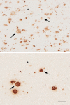
Adjacent cortical tissue sections from an AD patient, immunostained with antibodies R398 to Aβ42 (top) and R361 to Aβ40 (bottom). Two of the plaques that are present in both sections are denoted by arrows. Asterisks mark a blood vessel for reference. Bar = 100μm.
Figure 3
Figure 3
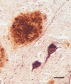
An Aβ plaque (brown) alongside intracellular tau tangles (purple) in the cortex of an AD patient. Combined polyclonal antibodies R398+R361 to Aβ40+42 plus monoclonal antibody CP13 to hyperphosphorylated tau. Bar = 20μm.
Figure 4
Figure 4
A neuritic Aβ plaque in consecutive sections of the cortex from an AD patient; The core is evident in the left-hand image, whereas sections through the periphery (middle and right) reveal only neurites (black). Serial sections may be required to unequivocally identify plaque types (a technical caveat noted by, among others, Alzheimer [1911] [reference 96]). Naoumenko-Feigin (silver) and periodic-Schiff stains. Bar = 20 μm for all images.
Figure 5
Figure 5
Variation in Aβ deposition in adjacent cortical gyri from an AD patient. Antibody 4G8, Nissl counterstain. Bar = 500μm.
Figure 6
Figure 6
Variable morphology of Aβ plaques in the cortex of an AD patient. Classical dense-cored plaques with the core-space-corona pattern are in the upper left and lower right, and an irregular cloud of diffuse material is near the center, along with numerous very small patches. Antibody 4G8; Nissl counterstain. Bar = 50μm.
Figure 7
Figure 7
The phases of Aβ plaque distribution in the brain [references 19, 145]; illustration courtesy of Dietmar Thal, KU Leuven.
Figure 8
Figure 8
Small, often stellate Aβ deposits in the cortex of an AD patient. Some Aβ accumulates within glial cells, most likely astrocytes (right). Antibody 4G8; Nissl counterstain. Bars = 20μm.
Figure 9
Figure 9
Band-like subpial Aβ (left) in neocortical layer 1 and presubicular lake-like Aβ (right) from two cases of AD. The subpial Aβ can be discontinuous, confluent, or punctate. Antibodies 4G8 (left) and 6E10 (right); Nissl counterstain. Bar = 100μm for both images.
Figure 10
Figure 10
Neurofibrillary tangles in the cortex of an AD patient immunostained with an antibody to Aβ40. When present, this colocalization occurs mostly on extracellular ('ghost') tangles. Nissl counterstain. Bar = 50μm.
Figure 11
Figure 11
Aβ deposits in white matter of an AD patient comprise clusters of small puncta and filamentous bundles. Left: Light-micrograph of a cluster immunolabeled with antibody 4G8 (Nissl counterstain). Right, electron micrograph of a punctum immunolabeled with antibody 4G8 (black dots are gold particles bound to the secondary antibody). Bars = 20μm (left) and 200nm (right).
Figure 12
Figure 12
Ultrastructure of fibrillar Aβ in the plaque corona (left) and core (right) in an AD patient. Bar = 500nm for both images.
Figure 13
Figure 13
High-magnification electron micrograph of a portion of the core of an Aβ-amyloid plaque in an AD patient. The fibrillarity of the material is less evident than in more peripheral zones. Unidentified particles (2 are marked by arrows) of various sizes and densities are interspersed among the amyloid fibrils; these can be found both in the core and corona. Bar = 200nm.
Figure 14
Figure 14
Aβ plaque with an antibody-refractory central core in an AD patient. Antibody 6E10; Nissl counterstain. Bar = 20μm.
Figure 15
Figure 15
Fluorescence-immunolabeled dyshoric cerebral Aβ-amyloid angiopathy (red; antibody R398) and tau-immunoreactive neurites (green; antibody CP13) in the cortex of an AD patient. Bar = 50μm.
Figure 16
Figure 16
Juxtavascular Aβ-plaque (arrow) in the cortex of an AD patient. Antibody 4G8, Nissl counterstain. Bar = 20μm.
Figure 17
Figure 17
Abnormal neurites associated with cortical Aβ plaques in two AD patients. Left: immunostain for neurofilament-H (antibody SMI31) with a Nissl counterstain; right, immunostain for a conformational epitope on tau filaments (antibody MC1). The presence of aberrant neurites that are immunoreactive for these antigens in plaques is variable. Bar = 25μm (right) and 50μm (left).
Figure 18
Figure 18
Abnormal neurite (top) containing organelles /debris adjacent to fibrillar amyloid (bottom) in the plaque corona of a patient with AD. Bar = 500nm.
Figure 19
Figure 19
Reactive astrocytes (left; antibody to GFAP) and microglia (right; antibody to IBA1) in cortical Aβ plaques of two AD patients. Despite some overlap of the two cell types within plaques, astrocytic somata tend to be more peripherally located than are microglial somata. Bar = 20μm for both panels.
Figure 20
Figure 20
Electron micrographs of a microglial cell in an Aβ-amyloid plaque of an AD patient. The white box in the image on the left denotes the region at higher magnification on the right. The fibrillar bundles of Aβ interdigitate with the microglial soma. Note that the microglial cytoplasm appears artifactually rarefied in this autopsy-derived tissue. Bar = 500 nm (right), 2.8μm (left).
Figure 21
Figure 21
Seeded Aβ deposition in the hippocampal formation of a TG2576 APP-transgenic mouse 5-months following unilateral injection of dilute AD brain extract into one hemisphere (left). The contralateral hippocampus in the same tissue section is on the right. Antibody 4G8; Bar = 100μm.
Figure 22
Figure 22
Aβ deposition (left) in the superior temporal gyrus and a neuritic plaque (right) in the hippocampal formation of two aged rhesus monkeys (Macaca mulatta; 35 years and ~30 years, respectively). Left: Antibody 82E1 to the N-terminal segment of Aβ, Nissl counterstain; Right: Antibody 06-17 to phosphorylated neurofilaments. Bars = 200μm (left) and 25μm (right). The maximum known lifespan of rhesus monkeys is 44 years (see Stonebarger et al. [2020] [reference 512]).
Figure 23
Figure 23
Aβ plaques in an aged (28 months) Tg2576 APP-transgenic mouse. Diffuse deposits are black, and some dense deposits have a golden core (one in the frontal cortex is magnified at right). Campbell-Gallyas silver stain. Bars = 1mm (left) and 50μm (right).

References

    1. Nelson, P.T., et al., Correlation of Alzheimer disease neuropathologic changes with cognitive status: a review of the literature. J Neuropathol Exp Neurol, 2012. 71(5): p. 362-81. - PMC - PubMed
    1. Glenner, G.G. and C.W. Wong, Alzheimer’s disease and Down’s syndrome: sharing of a unique cerebrovascular amyloid fibril protein. Biochem Biophys Res Commun, 1984. 122(3): p. 1131-5. - PubMed
    1. Glenner, G.G. and C.W. Wong, Alzheimer’s disease: initial report of the purification and characterization of a novel cerebrovascular amyloid protein. Biochem Biophys Res Commun, 1984. 120(3): p. 885-90. - PubMed
    1. Masters, C.L., et al., Amyloid plaque core protein in Alzheimer disease and Down syndrome. Proc Natl Acad Sci U S A, 1985. 82(12): p. 4245-9. - PMC - PubMed
    1. Masters, C.L. and K. Beyreuther, Pathways to the discovery of the Abeta amyloid of Alzheimer’s disease. J Alzheimers Dis, 2006. 9(3 Suppl): p. 155-61. - PubMed

LinkOut - more resources