Review
. 2021 Jan;17(3):e2005793.
doi: 10.1002/smll.202005793. Epub 2020 Dec 20.

Micro- and Nano-Devices for Studying Subcellular Biology

Affiliations
Review

Micro- and Nano-Devices for Studying Subcellular Biology

Michael J Siedlik et al. Small. 2021 Jan.

Abstract

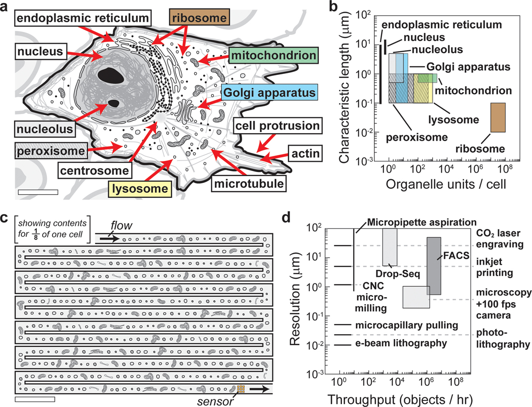
Cells are complex machines whose behaviors arise from their internal collection of dynamically interacting organelles, supramolecular complexes, and cytoplasmic chemicals. The current understanding of the nature by which subcellular biology produces cell-level behaviors is limited by the technological hurdle of measuring the large number (>103 ) of small-sized (<1 μm) heterogeneous organelles and subcellular structures found within each cell. In this review, the emergence of a suite of micro- and nano-technologies for studying intracellular biology on the scale of organelles is described. Devices that use microfluidic and microelectronic components for 1) extracting and isolating subcellular structures from cells and lysate; 2) analyzing the physiology of individual organelles; and 3) recreating subcellular assembly and functions in vitro, are described. The authors envision that the continued development of single organelle technologies and analyses will serve as a foundation for organelle systems biology and will allow new insight into fundamental and clinically relevant biological questions.

Keywords: devices; microelectronics; microfluidics; nanofluidics; organelles; subcellular structures.


Figures

Figure 1.
Figure 1.. Complexity and challenges in the analyses of subcellular materials due to the small size and large number of interacting subcellular components.
(a) Schematic representation of organelles and other subcellular structures within a cell. Scale bar, 5 μm. (b) Graphical representation of the size and number of organelles in a cell. (c) Schematic of a microfluidic channel for sensing organelles from cell lysate one at a time. The ~102 organelles depicted represent 1/8th of the mitochondria, peroxisomes, and lysosomes thought to reside in a typical cell and is intended to convey the challenge of analyzing the large number of small (sub-micrometer) structures found within each cell. Scale bar, 5 μm. (d) Graphical representation of the resolution and throughput of common techniques for biological analysis plotted with the resolution provided by common methods for fabricating micro and nano devices. See Supplementary Information for notes on the values used in (b) to estimate the characteristic size and quantity of each subcellular structure found in a cell, and in (d) to estimate the resolution and throughput of biological assays and fabrication techniques.
Figure 2.
Figure 2.. Micro and nano devices for the isolation and sorting of subcellular structures.
(a) Microfluidic systems enable organelles and cytoplasmic material to be harvested from defined regions of cells. (i) Schematic of a neuron cell culture system in which only axons grow through adjacent microchannels for subsequent lysing and harvesting for off-chip analysis. (ii) Images of GFP-expressing axons in this system before and after harvesting. Adapted with permission from Sgro et al.[35] Copyright 2013 American Chemical Society. (b) Micro devices immobilize organelles on-chip for subsequent imaging and other electrical measurements. (i) Schematic of a device designed to immobilize individual mitochondria by occluding their passage through nanoscale channels. (ii) Brightfield image of the device channels (from (i)) overlaid with the fluorescence image of individual mitochondria labeled with a fluorescent indicator of the organelle’s membrane potential. Scale bar, 10 μm. Adapted with permission from Zand et al.[47] Copyright 2013 American Chemical Society. (c) Devices sort organelles from unwanted cellular material based on size, electrical properties, or an optical signal. (i) Schematic of a device designed to sort vesicles by inducing downstream flow, based on the presence of a fluorescently labeled membrane protein, to direct the targets to a collection channel. Scale bar, 5 μm. (ii) Fluorescence images of a mixed population of vesicles without sorting (left) and the red population enriched following sorting (right). Adapted with permission from Schiro et al.[55] Copyright 2012 American Chemical Society.
Figure 3.
Figure 3.. Micro and nano devices for analyzing the composition of organelles and measuring their properties.
(a) Microfluidic devices encapsulate individual organelles within droplets for subsequent analyses. (i) Experimental workflow for sequencing RNA transcripts from individual nuclei using the Drop-Seq approach. (ii) Brightfield image of a microfluidic device encapsulating nuclei and beads into droplets. (iii) t-distributed stochastic neighbor visualization of >104 spinal cord nuclei. Reprinted from Sathyamurthy et al,[80] Copyright 2018, with permission from Elsevier. (b) Micro and nano devices can measure bioelectric properties of individual organelles. (i) Schematic of an electrofluidic device to measure the change in conductance as individual mitochondria flow over a carbon nanotube-based sensor. (ii) Experimental conductance measurements of flowing mitochondria (shown in red). Reprinted from Zand et al,[10] Copyright 2017, with permission from Elsevier.
Figure 4.
Figure 4.. Micro and nano devices for studying the function of subcellular units.
(a) Micro and nano devices provide well-defined conditions to visualize organelles and subcellular structures in vitro. (i) Brightfield image (left) and schematic (right) of a microfluidic system for encapsulating extract from fertilized Xenopus laevis eggs into cell-sized droplets to visualize the assembly of the mitotic spindle. (ii) Fluorescence image of a mitotic spindle (green: spindle poles, red: tubulin, blue: DNA) forming inside a droplet. From (i) Good et al, Science, 2013 [85] and (ii) Hazel et al, Science, 2013.[84] Reprinted with permission from AAAS. (b) Micro and nano devices enable control over temperature, reagents, and reaction volume for studying organelle assembly and disassembly. (i) Schematic of a microfluidic device for visualizing nucleolar assembly in a developing Drosophila embryo under rapidly changing temperature conditions. Inset: fluorescence image depicting the localization of the tagged nucleolar protein Fibrillarin (RFP-Fib) at different temperatures. (ii) Plots of the expected nucleolar assembly size in response to oscillating temperatures when assembly occurs via a phase transition or an ATP-dependent process, as well as experimental data for the assembly of two nucleolar proteins, Fibrillarin (RFP-Fib) and Modulo (eGFP-Mod). Adapted with permission from Falahati et al, 2017.[11] (c) Micro and nano devices produce synthetic organelles (SO) for uptake into living cells. (i) Schematic describing a SO designed to mimic the reactive oxygen species (ROS) processing capability of peroxisomes. (ii) Fluorescence images of menadione-stressed keratinocytes stained with CellRox Green to visualize intracellular ROS levels with and without the SOs. Reproduced with permission from Staufer et al, 2020.[90] Copyright 2020 The Authors.

References

    1. Alberts B, Johnson A, Lewis J, Raff M, Roberts K, Walter P, Molecular Biology of the Cell, Garland Science, New York, NY, 2008.
    1. Milo R, Jorgensen P, Moran U, Weber G, Springer M, Nucleic Acids Res. 2010, 38, D750. - PMC - PubMed
    1. Schier AF, Nat. Methods 2020, 17, 17. - PubMed
    1. Stubbington MJT, Rozenblatt-Rosen O, Regev A, Teichmann SA, Science (80-. ). 2017, 358, 58. - PMC - PubMed
    1. Eberwine J, Sul JY, Bartfai T, Kim J, Nat. Methods 2014, 11, 25. - PubMed

Publication types

LinkOut - more resources