Review
. 2021 May;159(5):1833-1842.
doi: 10.1016/j.chest.2020.12.007. Epub 2020 Dec 17.

Endobronchial Valves for the Treatment of Advanced Emphysema

Affiliations
Review

Endobronchial Valves for the Treatment of Advanced Emphysema

Karin Klooster et al. Chest. 2021 May.

Abstract

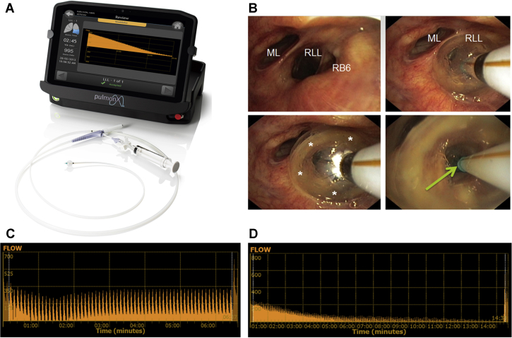
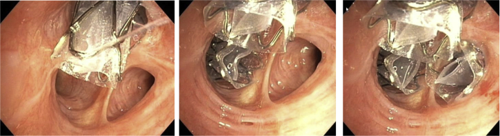
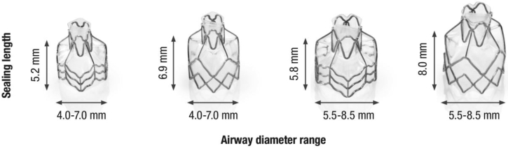
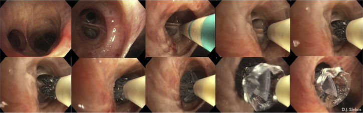
Bronchoscopic lung volume reduction with one-way endobronchial valves is a guideline treatment option for patients with advanced emphysema that is supported by extensive scientific data. Patients limited by severe hyperinflation, with a suitable emphysema treatment target lobe and with absence of collateral ventilation, are the responders to this treatment. Detailed patient selection, a professional treatment performance, and dedicated follow up of the valve treatment, including management of complications, are key ingredients to success. This treatment does not stand alone; it especially requires extensive knowledge of COPD for which the most appropriate treatment is discussed in a multidisciplinary approach. We discuss the endobronchial valve treatment for emphysema and provide a guideline for patient selection, treatment guidance, and practice tools, based on our own experience and literature.

Keywords: COPD; bronchoscopy; emphysema; endobronchial valve; lung volume reduction.


Figures

Figure 1
Figure 1
A-D, Case example of a suitable patient with COPD for bronchoscopic lung volume reduction with the use of one-way endobronchial valves. A, Inspiration chest CT scan shows lower lobe predominant heterogeneous emphysema; B, expiration chest CT scan shows predominant air trapping in the left lower lobe; C, perfusion scan confirms the left lower lobe target by absence of perfusion; D, quantitative CT analysis shows percentage of emphysematous destruction (at −950 Hounsfield Units), lobar volumes, and fissure integrity.
Figure 2
Figure 2
CT findings that exclude patients from endobronchial valve treatment. Each of these findings require a different treatment approach. (Reprinted with permission from Respiration, S. Karger AG, Basel, Switzerland).
Figure 3
Figure 3
A-C, Example of visual assessment approach of the high-resolution CT scan. A, Axial view; B, sagittal view; C, coronal view of 1 mm high-resolution CT scan reconstructions with the use of a sharp kernel. The red lines indicate the fissures. The right upper lobe shows more destruction compared with the other lobes. The fissures in the right lung look intact on all three views. The left major fissure looks intact in the axial view; however, in the coronal view, a small gap is visible. Based on these slices, with the use of a visual assessment, the right upper lobe is a potential Zephyr endobronchial valve (Zephyr EBV; Pulmonx Corporation, Redwood City, CA) target lobe. LLL = left lower lobe; LUL = left upper lobe; ML = middle lobe; RLL = right lower lobe; RUL = right upper lobe.
Figure 4
Figure 4
A-C, An example of quantitative analyses of high-resolution CT scan. A. Lung cartoon with per lobe the percentage of low attenuation areas (at −950 Hounsfield Units) values displayed. B. Rendering picture of the fissures. Green color indicates intact fissure; red color indicates a gap in the fissure. In this example, there is a gap visible in the left major fissure. This might imply that there is presence of collateral ventilation between left upper lobe and left lower lobe. C, Results of the percentage of fissure completeness, percentage of low attenuation areas (at −910 and −950 Hounsfield Units), and the inspiratory volumes are summarized per lobe. Based on this quantitative analysis (StratX Lung Report; Pulmonx Corporation, Redwood City, CA), the right upper lobe or the left upper lobe can be confirmed as potential targets for Zephyr endobronchial valve (Zephyr EBV; Pulmonx Corporation, Redwood City, CA). However, collateral ventilation needs to be ruled out by Chartis measurement. (Published with permission from Pulmonx Corporation). LLL = left lower lobe; LUL = left upper lobe; ML = middle lobe; RLL = right lower lobe; RML = right middle lobe; RUL = right upper lobe.
Figure 5
Figure 5
A-D, Chartis Pulmonary Assessment System. A, Chartis Pulmonary Assessment System (Pulmonx Corporation, Redwood City, CA) with Chartis balloon catheter. B, Example of temporary occlusion of the entrance of the right lower lobe with the Chartis balloon catheter. C, A continuous flow reading indicates collateral ventilation in the target lobe; no valves will be placed. D, A gradual decrease of the flow to no flow indicates the absence of collateral ventilation; valves can be placed into the target lobe. ML = middle lobe; RB6 = right lower lobe apical segment; RUL = right upper lobe.
Figure 6
Figure 6
Bronchoscopic view example of a right upper lobe treatment with endobronchial valves shows a full lobar occlusion to achieve the desired atelectasis of the lobe with consecutive valves placed in RB1, RB3, and RB2 (left to right).
Figure 7
Figure 7
The four available endobronchial valve sizes (Zephyr EBV; Pulmonx Corporation, Redwood City, CA).
Figure 8
Figure 8
Endobronchial valve placement example of the LB6 (apical segment of the left lower lobe). Clockwise, from left to right: left lower lobe entrance image, view of the LB6 with the Zephyr endobronchial valve (Zephyr EBV; Pulmonx Corporation, Redwood City, CA) catheter placed against the next subcarina indicates the length for a short size valve and subsequent deployment of the valve against this subcarina, which on further release expands against the LB6 wall, sealing it completely. The last picture shows the Zephyr endobronchial valve in place, with just the Heimlich valve part visible.

References

    1. Klooster K., ten Hacken N.H., Hartman J.E., Kerstjens H.A., van Rikxoort E.M., Slebos D.J. Endobronchial valves for emphysema without interlobar collateral ventilation. N Engl J Med. 2015;373(24):2325–2335. - PubMed
    1. Shah P.L., Slebos D.J. Bronchoscopic interventions for severe emphysema: where are we now? Respirology. 2020;25(9):972–980. - PubMed
    1. Welling J.B.A., Klooster K., Hartman J.E. Collateral ventilation measurement using chartis: procedural sedation vs general anesthesia. Chest. 2019;156(5):984–990. - PubMed
    1. Slebos D.J., Shah P.L., Herth F.J., Valipour A. Endobronchial valves for endoscopic lung volume reduction: best practice recommendations from expert panel on endoscopic lung volume reduction. Respiration. 2017;93(2):138–150. - PMC - PubMed
    1. Criner G.J., Sue R., Wright S. A multicenter randomized controlled trial of zephyr endobronchial valve treatment in heterogeneous emphysema (LIBERATE) Am J Respir Crit Care Med. 2018;198(9):1151–1164. - PubMed