. 2020 Dec 3:(166):10.3791/61545.
doi: 10.3791/61545.

Mitigation of Blood Borne Cell Attachment to Metal Implants through CD47-Derived Peptide Immobilization

Affiliations

Mitigation of Blood Borne Cell Attachment to Metal Implants through CD47-Derived Peptide Immobilization

Vaishali V Inamdar et al. J Vis Exp. .

Abstract

The key complications associated with bare metal stents and drug eluting stents are in-stent restenosis and late stent thrombosis, respectively. Thus, improving the biocompatibility of metal stents remains a significant challenge. The goal of this protocol is to describe a robust technique of metal surface modification by biologically active peptides to increase biocompatibility of blood contacting medical implants, including endovascular stents. CD47 is an immunological species-specific marker of self and has anti-inflammatory properties. Studies have shown that a 22 amino acid peptide corresponding to the Ig domain of CD47 in the extracellular region (pepCD47), has anti-inflammatory properties like the full-length protein. In vivo studies in rats, and ex vivo studies in rabbit and human blood experimental systems from our lab have demonstrated that pepCD47 immobilization on metals improves their biocompatibility by preventing inflammatory cell attachment and activation. This paper describes the step-by step protocol for the functionalization of metal surfaces and peptide attachment. The metal surfaces are modified using polyallylamine bisphosphate with latent thiol groups (PABT) followed by deprotection of thiols and amplification of thiol-reactive sites via reaction with polyethyleneimine installed with pyridyldithio groups (PEI-PDT). Finally, pepCD47, incorporating terminal cysteine residues connected to the core peptide sequence through a dual 8-amino-3,6-dioxa-octanoyl spacer, are attached to the metal surface via disulfide bonds. This methodology of peptide attachment to metal surface is efficient and relatively inexpensive and thus can be applied to improve biocompatibility of several metallic biomaterials.


Figures
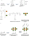
Figure 1:
Figure 1:. Schematic representation of the steps involved in appending pepCD47 to metal surfaces.
A) The clean metal samples are baked at 220 °C to oxidize the metal surface. The bisphosphonate groups of the polallylamine bisphosphonate with latent thiol groups (PABT) form co-ordinate bonds with the metal oxides and coat the metal surfaces to form a functionalized monolayer. The PABT coated metal surfaces are further treated with TCEP for deprotection of the thiol groups. B) The structure of PEI-PDT and symbolic representation. C) The PABT coated and TCEP reduced surfaces are treated with PEI-PDT which amplified the total number of thiol-reactive groups available for attachment of the thiolated peptide. Finally, PEI-PDT coated surfaces are reacted with terminal cysteine groups of pepCD47, and the peptide is attached to the surface via disulfide bonds.
Figure 2:
Figure 2:. Determining the immobilization density of pepCD47 on metal surface.
1cm X 1cm metal foils were modified using increasing concentrations (10, 30, 100 and 200 μg/mL) of fluorophore conjugated pepCD47. The excess peptide was removed using several washing steps then treated with 1mL TCEP solution to cleave the fluorophore conjugated fluorophore. The concentration of the peptide covalently attached to the metal surface was analyzed fluorimetrically using a standard curve prepared with a defined concentrations of fluorophore conjugated pepCD47. The immobilization density was represented as ng/cm2 of peptide attached to the metal surface. The data is expressed as mean ± SEM and is representative of at least three independent experiments.
Figure 3:
Figure 3:. Fluorescence microscopy imaging of the stainless steel surface modified with TAMRA-conjugated pepCD47.
Stainless steel mesh disks, consecutively modified with PABT, TCEP, PEI-PDT (A) or unmodified (B) were reacted with TAMRA-conjugated pepCD47. The properly conjugated and control bare metal meshes were extensively washed and imaged at 100x magnification. The scale bar length is 100 μm.
Figure 4:
Figure 4:. Evaluating acute anti-inflammatory and anti-thrombotic functions of pepCD47.
0.65 cm X 1cm metal foils were coated with 100 μg/mL of either human pepCD47 or scrambled peptide and exposed to blood in the Chandler loop apparatus. The unbound cells were removed by washing with PBS and the foils were fixed in 2% glutaraldehyde. The unmodified, scrambled modified and human pepCD47 modified surfaces were then incubated with the CFDA dye for 15 mins at 37 °C, washed with PBS and analyzed using a fluorescence microscope.
Figure 5:
Figure 5:. A prevalence of CD68-positive macrophages on the bare and pepCD47-functionalized metal surfaces.
Rat peripheral blood-derived monocytes were isolated by Ficoll gradient density centrifugation followed by negative immunoselection with magnetic microbeads. 5×10⁵ monocytes were added into the wells of a 12-well plate with individually placed bare metal foil samples (N=3) or the samples derivatized with rat pepCD47. Macrophage differentiation was stimulated by 100 ng/ml M-CSF. Six days after seeding the cells were fixed, and immunostained with anti-rat CD68 antibody, secondary Alexa Fluor-546 (red) conjugated antibody and counterstained with Hoechst 33342 nuclear dye (blue). Representative images of the bare metal (A) and pepCD47-functionalized (B) surfaces were captured at 200x magnification and merged. The scale bar length is 100 μm.

References

    1. Buccheri D, Piraino D, Andolina G & Cortese B Understanding and managing in-stent restenosis: a review of clinical data, from pathogenesis to treatment. Journal Thoracic Disease. 8 (10), E1150–e1162, (2016). - PMC - PubMed
    1. van Oeveren W Obstacles in haemocompatibility testing. Scientifica (Cairo). 2013. 392584, (2013). - PMC - PubMed
    1. Mitra AK & Agrawal DK In stent restenosis: bane of the stent era. Journal of Clinical Pathology. 59 (3), 232–239, (2006). - PMC - PubMed
    1. Iqbal J, Gunn J & Serruys PW Coronary stents: historical development, current status and future directions. British Medical Bulletin. 106 193–211, (2013). - PubMed
    1. Hoffmann R et al. Patterns and mechanisms of in-stent restenosis. A serial intravascular ultrasound study. Circulation. 94 (6), 1247–1254, (1996). - PubMed

Publication types

MeSH terms