Comparative Study
. 2020 Dec 21;18(12):e3001016.
doi: 10.1371/journal.pbio.3001016. eCollection 2020 Dec.

The SARS-CoV-2 Spike protein has a broad tropism for mammalian ACE2 proteins

Affiliations
Comparative Study

The SARS-CoV-2 Spike protein has a broad tropism for mammalian ACE2 proteins

Carina Conceicao et al. PLoS Biol. .

Abstract

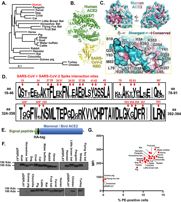
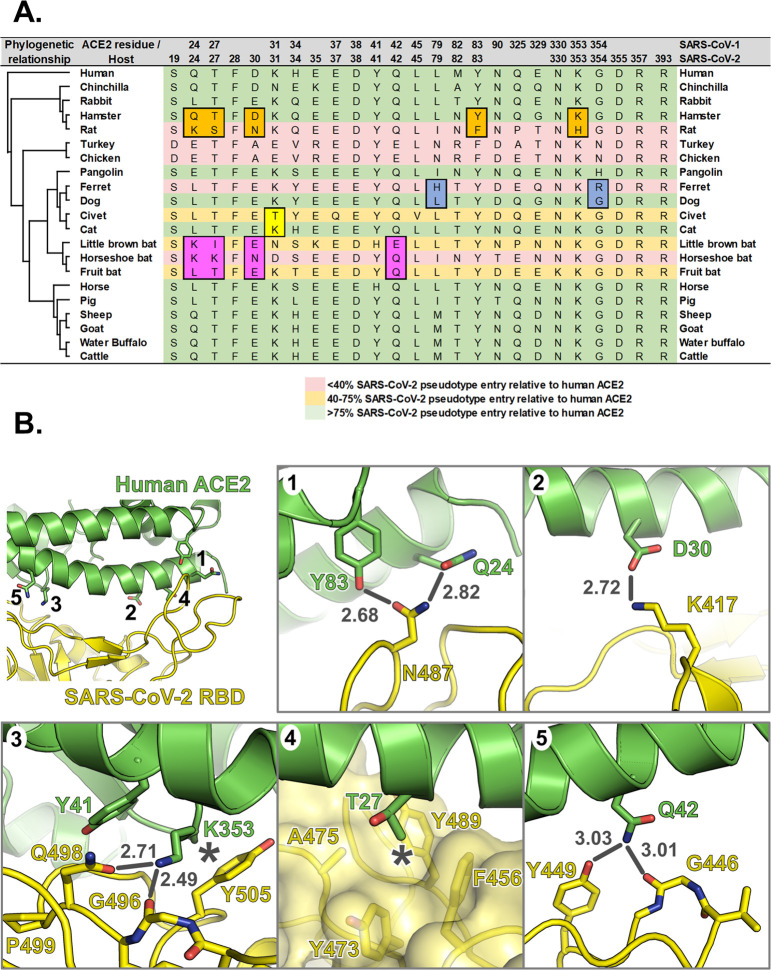
SARS Coronavirus 2 (SARS-CoV-2) emerged in late 2019, leading to the Coronavirus Disease 2019 (COVID-19) pandemic that continues to cause significant global mortality in human populations. Given its sequence similarity to SARS-CoV, as well as related coronaviruses circulating in bats, SARS-CoV-2 is thought to have originated in Chiroptera species in China. However, whether the virus spread directly to humans or through an intermediate host is currently unclear, as is the potential for this virus to infect companion animals, livestock, and wildlife that could act as viral reservoirs. Using a combination of surrogate entry assays and live virus, we demonstrate that, in addition to human angiotensin-converting enzyme 2 (ACE2), the Spike glycoprotein of SARS-CoV-2 has a broad host tropism for mammalian ACE2 receptors, despite divergence in the amino acids at the Spike receptor binding site on these proteins. Of the 22 different hosts we investigated, ACE2 proteins from dog, cat, and cattle were the most permissive to SARS-CoV-2, while bat and bird ACE2 proteins were the least efficiently used receptors. The absence of a significant tropism for any of the 3 genetically distinct bat ACE2 proteins we examined indicates that SARS-CoV-2 receptor usage likely shifted during zoonotic transmission from bats into people, possibly in an intermediate reservoir. Comparison of SARS-CoV-2 receptor usage to the related coronaviruses SARS-CoV and RaTG13 identified distinct tropisms, with the 2 human viruses being more closely aligned. Finally, using bioinformatics, structural data, and targeted mutagenesis, we identified amino acid residues within the Spike-ACE2 interface, which may have played a pivotal role in the emergence of SARS-CoV-2 in humans. The apparently broad tropism of SARS-CoV-2 at the point of viral entry confirms the potential risk of infection to a wide range of companion animals, livestock, and wildlife.


Conflict of interest statement

The authors have declared that no competing interests exist.

Figures

Fig 1
Fig 1. The SARS-CoV-2 binding site on ACE2 is highly variable.
(A) A phylogenetic tree of ACE2 proteins assembled using the neighbor-joining method [51] conducted in MEGA7 (Temple University, USA) [52] with ambiguous positions removed. The tree is drawn to scale, and support was provided with 500 bootstraps. (B) Structure of human ACE2 ectodomain (green) in complex with the RBD of SARS-CoV-2 [10]. (C) Conservation of mammalian ACE2 amino acid residues, estimated from site-specific evolutionary rates [50], mapped onto the surface of the ACE2 ectodomain [10], and coloured from blue (divergent) to purple (conserved) and presented in 2 orientations. Inset depicts the SARS-CoV-2 binding region of ACE2 (outlined), with residues that contact the SARS-CoV-2 RBD highlighted [6]. (D) WebLogo (University of California, Berkeley, USA) [53] plots summarising the amino acid divergence within the mammalian and bird ACE2 sequences characterised in this study. The single letter amino acid (aa) code is used with the vertical height of the amino acid representing its prevalence at each position in the polypeptide (aa 18–46, 78–91, 324–358, and 392–394 are indicated). The aa sites bound by SARS-CoV and SARS-CoV-2 Spike [11] are indicated by red arrows. *SARS-CoV-specific interactions. (E) ACE2 sequences were cloned into the pDISPLAY expression construct in frame with an N-terminal signal peptide (the murine Ig κ-chain leader sequence) and HA-tag. (F) Expression of individual mammal or bird ACE2 proteins was confirmed at a whole cell level by western blot. (G) Flow cytometry was performed to examine surface expression of each ACE2 protein on non-permeabilised cells. For gated cells, the percentage positivity and MFI are plotted. The data underlying this figure may be found in S1 Data and S1 Raw Images. aa, amino acid; ACE2, angiotensin-converting enzyme 2; MFI, mean fluorescence intensity; RBD, receptor binding domain; SARS-CoV, SARS Coronavirus; SARS-CoV-2, SARS Coronavirus 2.
Fig 2
Fig 2. Receptor screening using surrogate entry assays identifies SARS-CoV-2 Spike as a pan-tropic viral attachment protein.
(A) A heatmap illustrating the receptor usage profile of SARS-CoV-2 and SARS-CoV in pseudotype entry and cell–cell fusion assays with various mammalian and bird ACE2s. The data in each row are normalised to the signal seen for human ACE2 (top), with results representing the mean percentage calculated from 3 separate experiments performed on different days. A vector-only control (pDISPLAY) was added to demonstrate specificity. Mammalian and bird ACE2s are organised, top to bottom, based on their phylogenetic relationship (rectangular cladogram, left). The inter-experimental standard error of the mean for the pseudotype and cell–cell fusion assays ranged from 0.01% to 47.92% (median 10.73%) and 0.12% to 32.97% (median 5.43%), respectively. (B and C) For both SARS-CoV-2 and SARS-CoV, the respective cell–cell and pseudotype assay percentages for each ACE2 protein (relative to human ACE2) were plotted on an XY scatter graph, the Pearson correlation calculated and a linear line of regression fitted together with 95% confidence intervals. The data underlying this figure may be found in S1 Data. ACE2, angiotensin-converting enzyme 2; SARS-CoV, SARS Coronavirus; SARS-CoV-2, SARS Coronavirus 2.
Fig 3
Fig 3. A cognate ACE2 receptor is required for SARS-CoV-2 infection.
(A) Various cell lines derived from birds, dogs, rabbits, rodents, pigs, ruminants, and primates were experimentally infected with SARS-CoV-2 at a MOI of 0.001. At 72 h postinfection, the supernatants from cells were harvested and titred by TCID-50. For each cell line, RNA from uninfected cells was also extracted, and RT-qPCR was performed to detect ACE2 mRNA, with the value above each line indicating the cycle when PCR positivity was achieved (Ct). (B) Four of the same cell lines were infected again, this time at high MOI (1). (C) BHK-21 hamster cells were transiently transfected with ACE2 expression constructs (or a vector control [pDISPLAY]) before being infected with SARS-CoV-2 at high MOI (1). *p < 0.05 Student t test, compared with pDISPLAY. For all high MOI experiments, supernatant samples were harvested at 48 hpi for titration by TCID-50. The DL for the TCID-50 is indicated. In all experiments, the initial inoculum used for infection was titred, and infections were performed in duplicate, with error bars denoting standard deviation from the mean. The data underlying this figure may be found in S1 Data. ACE2, angiotensin-converting enzyme 2; BHK-21, baby hamster kidney 21; Ct, cycle threshold; DL, detection limit; MOI, multiplicity of infection; RT-qPCR, reverse transcription quantitative PCR; SARS-CoV-2, SARS Coronavirus 2; TCID50, 50% tissue culture infective dose.
Fig 4
Fig 4. Amino acid substitutions within SARS-CoV-2 Spike RBD may have contributed to zoonotic emergence.
(A) Comparison of the RBD structures of SARS-CoV-2 and RaTG13 Spike proteins [6,12] identified a high degree of structural similarity. Nevertheless, a number of amino acid changes between RaTG13 and SARS-CoV-2 were identified at residues interacting directly with ACE2 (according to [6]). SARS-CoV-2 N439, which does not interact directly with ACE2, was included because of its functional stabilisation role in the 498–505 loop, its previous identification within the SARS-CoV Spike RBD–ACE2 interface [19], and the N439K substitution present in RaTG13. (B) An XY scatter plot demonstrating the receptor usage profile of RaTG13 Spike in pseudotype (X) and cell–cell fusion assays (Y). Each point represents the mean signal seen from 3 experiments performed on separate days, with the human ACE2 highlighted in blue. Human ACE2 utilisation by SARS-CoV-2 and SARS-CoV Spike is also plotted for reference. In this graph, values are not normalised to human ACE2 since RaTG13 is, to our knowledge, not a human-tropic virus. (C and D) Specific amino acids within the RBDs of SARS-CoV-2 and RaTG13 Spike, which directly interact with ACE2 yet vary between these 2 sequences, were mutated to generate chimaeric SARS-CoV-2-RaTG13 Spike proteins. The cell–cell fusion activity of individual point mutants, as well as a full chimaera containing all 8 mutations (chimaera), was then examined using human ACE2 expressing target cells with activity normalised to the fusion seen with the wildtype (WT) viral glycoprotein of (C) SARS-CoV-2 or (D) RaTG13. The data points shown are the mean cell–cell fusion results seen in 3 completely independent experiments (green, increased relative activity; red, decreased relative activity). *p < 0.05 Student t test, compared with WT. (E) Expression of the same mutants was analysed by western blot targeting the flag tag fused to these Spike proteins (S, full-length Spike; S1/S2, cleaved variant). Results are representative of protein expression experiments performed in duplicate, with a GAPDH loading control also shown. The data underlying this figure may be found in S1 Data and S1 Raw Images. ACE2, angiotensin-converting enzyme 2; GAPDH, glyceraldehyde 3-phosphate dehydrogenase; RBD, receptor binding domain; RLU, relative light units; SARS-CoV, SARS Coronavirus; SARS-CoV-2, SARS Coronavirus 2; WT, wildtype.
Fig 5
Fig 5. Substitutions at the interface between SARS-CoV-2 RBD and mammalian ACE2 proteins impact receptor utilisation.
(A) Residues of mammalian ACE2 sequences used in this study that are predicted to interact with the RBD SARS-CoV and SARS-CoV-2, based on the structures of human ACE2 in complex with SARS-CoV [19] and SARS-CoV-2 [6]. Differences between closely related species that may impact RBD binding are highlighted. (B) Interface between human ACE2 (green) and SARS-CoV-2 RBD (yellow). Insets 1 to 5 show molecular interactions discussed in the main text. Bonds that may be disrupted are shown as grey lines, with bond distances in grey text, and hydrophobic interactions that may be disrupted are marked with asterisks. ACE2, angiotensin-converting enzyme 2; RBD, receptor binding domain; SARS-CoV, SARS Coronavirus; SARS-CoV-2, SARS Coronavirus 2.
Fig 6
Fig 6. Amino acid variation in SARS-CoV-2 and RaTG13 RBD impacts human ACE2 receptor utilisation.
Structure of RaTG13 RBD (orange) [12] superposed onto the structure of human ACE2 (green) in complex with the SARS-CoV-2 RBD (yellow) [10]. Selected RBD residues that promote association with ACE2 are highlighted, as are the ACE2 “hotspot” residues K31 and K353 [13]. Insets 1 to 5 show molecular interactions discussed in the main text. Bonds that may be disrupted are shown as grey lines, with bond distances in grey text, and hydrophobic interactions that may be disrupted or potential steric clashes are marked with asterisks. ACE2, angiotensin-converting enzyme 2; RBD, receptor binding domain; SARS-CoV-2, SARS Coronavirus 2.

References

    1. Zhou P, Yang XL, Wang XG, Hu B, Zhang L, Zhang W, et al. A pneumonia outbreak associated with a new coronavirus of probable bat origin. Nature. 2020;579 (7798):270–3. 10.1038/s41586-020-2012-7 - DOI - PMC - PubMed
    1. Li W, Shi Z, Yu M, Ren W, Smith C, Epstein JH, et al. Bats are natural reservoirs of SARS-like coronaviruses. Science. 2005;310 (5748):676–9. 10.1126/science.1118391 - DOI - PubMed
    1. Azhar EI, El-Kafrawy SA, Farraj SA, Hassan AM, Al-Saeed MS, Hashem AM, et al. Evidence for camel-to-human transmission of MERS coronavirus. N Engl J Med. 2014;370 (26):2499–505. 10.1056/NEJMoa1401505 - DOI - PubMed
    1. Huang A.T., Garcia-Carreras B., Hitchings M.D.T., Yang B., Katzelnick L., Rattigan S.M., Borgert B., Moreno C., Solomon B.D., Rodriguez-Barraquer I., Lessler J., Salje H., Burke D.S., Wesolowski A., and Cummings D.A.T., A systematic review of antibody mediated immunity to coronaviruses: antibody kinetics, correlates of protection, and association of antibody responses with severity of disease. medRxiv, 2020. - PMC - PubMed
    1. Shang J, Ye G, Shi K, Wan Y, Luo C, Aihara H, et al. Structural basis of receptor recognition by SARS-CoV-2. Nature. 2020;581 (7807):221–4. 10.1038/s41586-020-2179-y - DOI - PMC - PubMed

Publication types

Substances