. 2020 Dec 21;16(12):e1009176.
doi: 10.1371/journal.ppat.1009176. eCollection 2020 Dec.

GAS6 signaling tempers Th17 development in patients with multiple sclerosis and helminth infection

Affiliations

GAS6 signaling tempers Th17 development in patients with multiple sclerosis and helminth infection

Juan M Ortiz Wilczyñski et al. PLoS Pathog. .

Abstract

Multiple sclerosis (MS) is a highly disabling neurodegenerative autoimmune condition in which an unbalanced immune response plays a critical role. Although the mechanisms remain poorly defined, helminth infections are known to modulate the severity and progression of chronic inflammatory diseases. The tyrosine kinase receptors TYRO3, AXL, and MERTK (TAM) have been described as inhibitors of the immune response in various inflammatory settings. We show here that patients with concurrent natural helminth infections and MS condition (HIMS) had an increased expression of the negative regulatory TAM receptors in antigen-presenting cells and their agonist GAS6 in circulating CD11bhigh and CD4+ T cells compared to patients with only MS. The Th17 subset was reduced in patients with HIMS with a subsequent downregulation of its pathogenic genetic program. Moreover, these CD4+ T cells promoted lower levels of the co-stimulatory molecules CD80, CD86, and CD40 on dendritic cells compared with CD4+ T cells from patients with MS, an effect that was GAS6-dependent. IL-10+ cells from patients with HIMS showed higher GAS6 expression levels than Th17 cells, and inhibition of phosphatidylserine/GAS6 binding led to an expansion of Th17 effector genes. The addition of GAS6 on activated CD4+ T cells from patients with MS restrains the Th17 gene expression signature. This cohort of patients with HIMS unravels a promising regulatory mechanism to dampen the Th17 inflammatory response in autoimmunity.


Conflict of interest statement

The authors have declared that no competing interests exist.

Figures

Fig 1
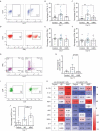
Fig 1. Helminth infection differentially modulates TAM receptor and their ligands expression in patients with MS and type 2 environment.
A) Representative dot plots showing the three main sub-populations of mononuclear cells based on the expression of CD11b, CD4, and CD1c. TYRO3, AXL, and MERTK expression were evaluated in circulating monocytes (CD11bhighCD4mid) and DCs (CD1chighCD11blow) of patients with MS, patients with HIMS, and healthy controls by flow cytometry. B-G) The percentage of TYRO3-positive as well as the mean fluorescent intensity (MFI) of the receptor on monocytes (B, C, and D) and DCs (E, F, and G) are graphed. H, I, and J) The percentage of AXLhigh as well as the MFI of the receptor on DCs are shown. K, L, and M) The expression of MERTK on DCs is shown as both percentage and MFI. N, O, and P) The percentage of circulating CD4+ T cell expressing GAS6 as well as its MFI are shown. Q, R, and S) The percentage of circulating CD4+ T cell expressing PROS1 and its MFI are shown. Data is presented as a pool of independent samples included in the specific staining (Control N = 21–31; MS N = 10–27; and HIMS N = 11–16). One-way ANOVA with a Fisher post hoc test was performed to determine statistical significances, *p<0.05 **p≤0.01 ****p≤0.001. MS = multiple sclerosis, HIMS = helminth-infected multiple sclerosis.
Fig 2
Fig 2. Concomitant helminth infection induced a significantly lower percentage of Th17 cells and transcriptional program in patients with MS.
A) Representative dot plot showing CD4+IL-17+ cells, based on the isotype control gating on the left. B and C) The percentage of double positive cells at day 7 and 10, post activation with anti-CD3/CD28, are graphed. D) Representative dot plot showing CD4+IFNγ+ gate, based on the isotype control. E and F) The percentage of double positive cells after 7, and 10 days post-stimulation, are shown. G and H) Representative dot plot and the percentage of IL-17+IFNγ+ double positive in CD4+ T cells at day 10 after stimulation in the three clinical groups are shown. I and J) Representative dot plot showing the percentage of CD4+IL-4+ T cells, based on the isotype control, after 17 days post-stimulation, and the independent samples analyzed. K) Heat map showing relative mRNA expression profile of Th17 lineage-specific and related genes in stimulated vs non-stimulated T cells of MS and HIMS cohorts. All relative expression referenced the non-stimulated healthy control for basal expression levels (equal 0). The expression levels of indicated gene were evaluated by qPCR and referred as 2^dCT. EF1A1 was used as the housekeeping gene. The numbers in the heat map indicate the mean value of at least 4 independent samples. Data is presented as a pool of independent samples included in the specific assay (Control N = 4–23; MS N = 6–22; and HIMS N = 4–12). One-way ANOVA with a Fisher post hoc test was performed to determine statistical significance, *p<0.05 **p≤0.01 ****p≤0.001. MS = multiple sclerosis, HIMS = helminth-infected multiple sclerosis.
Fig 3
Fig 3. Factor analysis of mixed data shows that the differential expression of TAM components explains and contributes to the segregation of both cohorts of patients.
To analyze the similarity and relationship among 24 variables studied in both MS and HIMS cohorts, we have performed the factor analysis of mixed data (FAMD) dimensionality reduction approach. We have included TAM receptors and ligands expression in each leukocyte population (CD11bhigh, CD1chigh, and CD4+), the percentage of T-helper cells, score disease (EDSS), age, eosinophils, IgE titers, and total leukocyte and lymphocyte numbers. A) Individual Factor Map shows the distribution of the two most informative dimensions (Dim1 and Dim2) that explain the 39.4% of variance. Patients from MS group were highlighted in light-blue (1–29) and HIMS cohort (30–47) in purple for visualization. B) The bar graph indicates the top 20 individual variables that contribute to segregate Dim 1. The red dashed line indicates the expected average contribution. Variables over the cut-off would be considered as important contributors. C) Hierarchical clustering analysis shows that the cluster n°3 separately groups most of HIMS patients while cluster n°2 groups the majority of MS patients. The arrowheads show some patients of MS (5 and 11) or HIMS (31 and 39) clustering differentially.
Fig 4
Fig 4. CD4+ T cells from patients with MS and concomitant helminth infection induced lower DCs activation that is GAS6-dependent.
Mixed lymphocyte reaction (MLR) assay was performed by co-culturing monocyte-derived DCs from healthy controls with heterologous sorted CD4+ T cells from patients with MS and HIMS at a 1:5 ratio for 72 hours. Activation status of DC was evaluated by measuring surface levels of co-stimulatory molecules. A, B, and C) Relative expression levels of CD80 (A), CD86 (B), and CD40 (C) on CD11c+ DC cells after MLR assay. Expression levels were calculated relative to basal expression of DCs alone. D, E, and F) Blocking antibody against GAS6 (2 μg/mL) or its corresponding isotype was added in the MLR assays, and relative expression levels of CD80 (D), CD86 (E), and CD40 (F) on CD11c+ cells was calculated. The MLR assay was assessed employing 4–7 independent monocyte-derived DCs co-cultured with sorted CD4+ T cells from at least 4–6 different patients of each clinical group. Paired t-test was performed for each activation marker and statistical significances are indicated as *p<0.05 **p≤0.01. MS = multiple sclerosis, HIMS = helminth-infected multiple sclerosis.
Fig 5
Fig 5. GAS6 expression is higher in CD4+ IL-10+ cells than in CD4+IL17+ cells and modulates Th17 development in HIMS.
A and B) Expression levels of PROS1 and GAS6 in CD4+ IL-17-expressing cells, measured as MFI, are shown. Independent data for each specific staining are shown, and the negative threshold is indicated as a black dash line. The baseline expression of non-stimulated CD4+ T cells is indicated as a blue dash line. C and D) The percentage of CD4+IL10+ cells was positively correlated with the percentage of Th17 subset. E) Comparison of GAS6 expression levels between IL-10+ and IL17+ CD4+ T cells within each clinical group. F-I) Sorted CD4+ T cells from healthy controls were activated and expanded with anti-CD3/CD28 during 5 days in the presence of AnnV (1 μg/mL), a competitor for phosphatidylserine binding with TAM ligands, or blocking anti-GAS6 (2 μg/mL) antibody; IL-17 and IFNγ mRNA expression were evaluated by quantitative PCR (qPCR). J) Transcription factor genes related to the Th17 subset (IRF4, LXRα, cMAF, AHR and HIF1α) were also evaluated in stimulated CD4+ T cells under anti-GAS6 treatment by qPCR. K and L) mRNA level of IL-17 and IFNγ were evaluated in activated CD4+ T cells from HIMS group after blocking GAS6. M-S) CD4+ T cells from controls and patients with MS were activated with anti-CD3/CD28 and treated with 50 nM of recombinant human GAS6 every 2 days during 5 days and IL-17 and IFNγ, IL-22, cMAF, SGK1, HIF1α, and AHR were evaluated by qPCR. Correlation was assessed by Spearman test. Flow cytometry data is presented as a pool of independent samples included in the specific staining (Control N = 4–14; MS = 6–15; HIMS = 6–10). qPCR was performed with at least 5 independent HC, 3 of MS, and 3 of HIMS donors. One-way ANOVA with a Fisher post hoc test was used in A-D and statistical significances are indicated as *p<0.05 **p≤0.01. One-tailed paired t-test was used to compare blocking conditions. MS = multiple sclerosis, HIMS = helminth-infected multiple sclerosis.
Fig 6
Fig 6. Helminth-induced type 2 immunity enhances regulatory TAM/GAS6 signaling to dampen Th17 pathological responses in multiple sclerosis.
The host type 2 immune response is elicited to clear parasitic helminth infections; however, the co-evolution of these parasites has promoted a variety of mechanisms to oppose and redirect immune responses by manipulating immune cell programming and function. Integrating our results with the current knowledge, we propose that gastrointestinal (1) helminths infections can enhance the regulatory axis of TAM receptors (TYRO3, AXL, and MERTK) in peripheral blood (2) CD11bhigh and CD1chigh cells, and their ligand GAS6 in CD4+ T cells of patients with MS. This negative regulatory pathway could be essential for dampening co-stimulatory signals (CD40, CD80, and CD86) of antigen-presenting cells and controlling the pathological Th17 (pTh17) response at secondary lymphoid organs (3). The active chronic infection resets T helper response toward Th2/regulatory signals, limiting pathological Th17 (pTh17) effector response. This reprogramming of CD4+ T cells and GAS6 signaling under a helminth-induced type 2 environment could be essential for maintaining the balance between CD4+ IL-10+ and Th17 cells and reducing inflammatory IL-17 and IFNγ signals in the central nervous system (4). In summary, GAS6 tempers not only the innate immune response but also regulates Th17 development in an autocrine/paracrine manner.

Similar articles

Cited by

References

    1. Baecher-Allan C, Kaskow BJ, Weiner HL. Multiple Sclerosis: Mechanisms and Immunotherapy. Neuron. 2018;97:742–68. 10.1016/j.neuron.2018.01.021 - DOI - PubMed
    1. Sospedra M, Martin R. Immunology of Multiple Sclerosis. Semin Neurol. 2016;36:115–27. 10.1055/s-0036-1579739 - DOI - PubMed
    1. Dendrou CA, Fugger L, Friese MA. Immunopathology of multiple sclerosis. Nat Rev Immunol. 2015;15:545–58. 10.1038/nri3871 - DOI - PubMed
    1. Montes M, Zhang X, Berthelot L, Laplaud DA, Brouard S, Jin J, et al. Oligoclonal myelin-reactive T-cell infiltrates derived from multiple sclerosis lesions are enriched in Th17 cells. Clin Immunol. 2009;130:133–44. 10.1016/j.clim.2008.08.030 - DOI - PMC - PubMed
    1. Greter M, Heppner FL, Lemos MP, Odermatt BM, Goebels N, Laufer T, et al. Dendritic cells permit immune invasion of the CNS in an animal model of multiple sclerosis. Nat Med. 2005;11:328–34. 10.1038/nm1197 - DOI - PubMed

Publication types

Substances