The effect of platelet-rich fibrin (PRF) on maxillary incisor retraction rate
- PMID: 33347530
- PMCID: PMC8028489
- DOI: 10.2319/050820-412.1
The effect of platelet-rich fibrin (PRF) on maxillary incisor retraction rate
Abstract
Objective: To investigate the efficiency of platelet-rich fibrin (PRF) injection on maxillary incisor retraction rate.
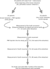
Materials and methods: The study included 40 patients (23 women and 17 men; mean age; 20.7 ± 1.45) with Class II Division 1 malocclusion. The treatment plan for all patients was extraction of the maxillary first premolars and canine distalization, followed by retraction of the maxillary incisors. Patients were randomly divided into two groups. The study group received injectable platelet-rich fibrin (i-PRF) two times with an interval of 2 weeks; the control group did not receive i-PRF. In both groups, the measurements were bilaterally assessed as the distances between the lateral and canine teeth on the plaster models at five time points. The rate of incisor movement was evaluated by Student's t-test, analysis of variance, and Tukey honestly significant difference tests. Statistical significance was set as P < .05.
Results: The average movements of incisors were significantly higher in the study group than the control group at all time points (P < .05). According to the within-group comparison, none of the measurements showed any significant differences between the right and left sides in both groups at all time points (P > .05). While the movement of incisors was significantly higher in the study group in the week following the PRF injection compared to the other weeks (P < .05), there were no significant differences in the control group at all-time points (P > .05).
Conclusions: Applying i-PRF significantly increased the rate of maxillary incisor retraction at all time intervals. Platelet-rich fibrin injection can be an effective method for shortening treatment duration.
Keywords: Incisor retraction rate; Injectable platelet-rich fibrin; Rate of tooth movement; i-PRF.
© 2021 by The EH Angle Education and Research Foundation, Inc.
Figures


Comment in
-
Response to the Letter.Angle Orthod. 2021 Nov 1;91(6):859. doi: 10.2319/0003-3219-91.6.859. Angle Orthod. 2021. PMID: 34670270 Free PMC article. No abstract available.
-
Letter to the Editor.Angle Orthod. 2021 Nov 1;91(6):858. doi: 10.2319/0003-3219-91.6.858. Angle Orthod. 2021. PMID: 34670271 Free PMC article. No abstract available.
-
Response to the Letter.Angle Orthod. 2022 Mar 1;92(2):295. doi: 10.2319/1945-7103-92.2.295. Angle Orthod. 2022. PMID: 35168263 Free PMC article. No abstract available.
-
Letter to the Editor.Angle Orthod. 2022 Mar 1;92(2):294. doi: 10.2319/1945-7103-92.2.294. Angle Orthod. 2022. PMID: 35168264 Free PMC article. No abstract available.
References
-
- Fink DF, Smith RJ. The duration of orthodontic treatment. Am J Orthod Dentofacial Orthop. 1992;102:45–51. - PubMed
-
- Alikhani M, Chou MY, Khoo E, et al. Age-dependent biologic response to orthodontic forces. Am J Orthod Dentofacial Orthop. 2018;153:632–644. - PubMed
-
- Skidmore KJ, Brook KJ, Thomson WM, Harding WJ. Factors influencing treatment time in orthodontic patients. Am J Orthod Dentofacial Orthop. 2006;129:230–238. - PubMed
-
- Bishara SE, Ostby AW. White spot lesions: formation, prevention, and treatment. Semin Orthod. 2008;14:174–182.
Publication types
MeSH terms
LinkOut - more resources
Full Text Sources

