Review
. 2020 Dec;239(3):189-208.
doi: 10.1086/711293. Epub 2020 Nov 20.

Dopamine as a Multifunctional Neurotransmitter in Gastropod Molluscs: An Evolutionary Hypothesis

Review

Dopamine as a Multifunctional Neurotransmitter in Gastropod Molluscs: An Evolutionary Hypothesis

Mark W Miller. Biol Bull. 2020 Dec.

Abstract

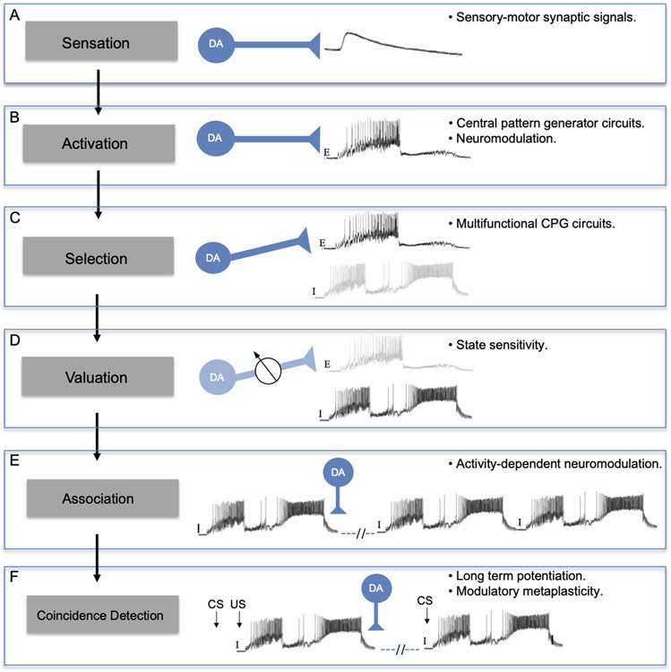
AbstractThe catecholamine 3,4-dihydroxyphenethylamine, or dopamine, acts as a neurotransmitter across a broad phylogenetic spectrum. Functions attributed to dopamine in the mammalian brain include regulation of motor circuits, valuation of sensory stimuli, and mediation of reward or reinforcement signals. Considerable evidence also supports a neurotransmitter role for dopamine in gastropod molluscs, and there is growing appreciation for its potential common functions across phylogeny. This article reviews evidence for dopamine's transmitter role in the nervous systems of gastropods. The functional properties of identified dopaminergic neurons in well-characterized neural circuits suggest a hypothetical incremental sequence by which dopamine accumulated its diverse roles. The successive acquisition of dopamine functions is proposed in the context of gastropod feeding behavior: (1) sensation of potential nutrients, (2) activation of motor circuits, (3) selection of motor patterns from multifunctional circuits, (4) valuation of sensory stimuli with reference to internal state, (5) association of motor programs with their outcomes, and (6) coincidence detection between sensory stimuli and their consequences. At each stage of this sequence, it is proposed that existing functions of dopaminergic neurons favored their recruitment to fulfill additional information processing demands. Common functions of dopamine in other intensively studied groups, ranging from mammals and insects to nematodes, suggest an ancient origin for this progression.


Figures

Figure 1.
Figure 1.
TH-like immunoreactivity in the albumen gland of the opisthobranch Bursatella leachii. (A) Low power image showing bundles of THli fibers (arrow) entering the albumen gland. Small immunoreactive somata were observed adhering to the fiber tracts. Fine axons (arrowheads) covered the entire surface of the gland. Calibration bar = 200 μm. (B) At higher magnification, some of the small (5 – 10 μm diameter) THli cell bodies were solitary, and others were clustered into small groups (arrowheads). Branch point of the fiber bundle (arrow) corresponds to arrow in panel A. Calibration bar = 50 μm. (Unpublished data; G. Rosado-Mattei and M.W. Miller, Institute of Neurobiology, University of Puerto Rico).
Figure 2.
Figure 2.
Dopamine as a sensory neurotransmitter in gastropods. (A) Low magnification image shows abundant TH-like immunoreactive innervation of the lips in Biomphalaria glabrata. Scale bar = 100 μm. (B) Higher magnification of THli cells in the mantle integument of Biomphalaria alexandrina. A single process from each cell projects through the epithelium, terminating as a bulbous enlargement at the surface (arrows). Scale bar = 20 μm. A, B: Reprinted with permission from Vallejo et al., J. Comp. Neurol. 522: 2532-2552, 2014. (C) THli cells in the oral veil of Pleurobranchaea californica. Groups of cilia-like terminations (arrows) penetrate the epithelial surface. Scale bar = 20 μm. Reprinted with permission from Brown et al., PLoS ONE 13: e0208891, 2018. (D) Esophageal nerve tracing (biocytin, red) and THli (green) on the surface of the pharynx of Aplysia californica. One double-labeled cell (yellow, arrow) in this merged image is a THli neuron that projects toward the CNS via the esophageal nerve. Scale bar = 50 μm. Reprinted with permission from Martínez-Rubio et al., J. Comp. Neurol. 514: 329-342, 2009.
Figure 3.
Figure 3.
The giant dopaminergic pedal neuron of Biomphalaria glabrata. (A) Intracellular recording from LPeD1 (lower trace) of B. glabrata; isolated CNS in normal saline solution. The recording exhibits alternating phases of activity and quiescence. A putative cardio-respiratory motor neuron in the visceral ganglion (VJ Mn, upper trace) was observed to burst out of phase with LPeD1. Calibration bars: 5 s, 20 mV. (B) Changing the bathing medium to a raised divalent saline solution (Hi-Di) eliminated polysynaptic signaling, enabling resolution of direct one-to-one inhibitory postsynaptic potential (IPSPs, upper record). Each IPSP occurred with a brief and constant latency following each LPeD1 impulse (lower record). Calibration bars = 1 s; 5 mV, upper record; 20 mV, lower record. (C) Pharmacological evidence for direct dopaminergic signaling from LPeD1 to VJ Mn. Control: Depolarizing current pulses were passed into LPeD1 producing sequentially greater number of impulses (lower record). The IPSPs produced in the VJ Mn became progressively larger as more impulses were stimulated. When the dopaminergic (D2) antagonist sulpiride was added to the solution (100 uM), the IPSPs were blocked. They returned following approximately 15 min Wash (right panel). Calibration bars = 0.5 s, 20 mV, 20 mV. (D-F) Morphological confirmation of LPeD1 and VJ Mn identity. (D) Neurobiotin was injected into LPeD1, a second neighboring cell in the pedal ganglion, and the VJ Mn. The three injected neurons were visualized with Avidin 546 (false color magenta). The axon of LPeD1 can be seen descending through the left pleural ganglion (arrow), while the neighboring cell projects to the contralateral pedal ganglion (arrowhead). (E) When the preparation was processed for TH-like immunoreactivity and viewed with avidin 488 (green), only the LPeD1 neuron and its descending axon (arrow) were labeled. (F) When the fill (D) and immunohistochemical (E) panels were overlaid, only the LPeD1 neuron and its descending axon (arrow) appear white (colocalization). The other injected cells, VJ Mn and the neighboring pedal neuron (arrowhead), appear magenta. Calibration bar = 50 μm, applies to D-F. Reprinted from Vallejo et al., 2014, J. Comp. Neurol. 522: 2532-2552, with permission from John Wiley & Sons, Inc.
Figure 4.
Figure 4.
Dopamine activates central pattern generator circuits of gastropods. (A) The dopaminergic RPeD1 interneuron of Lymnaea stagnalis was co-cultured with interneurons Ip3I and VD4 for 24 h, allowing the three cells to extend neurites and form synapses. Passing hyperpolarizing current into RPeD1 (asterisk) established that there was no electrical coupling between the cells. Passing depolarizing current that caused RPeD1 to fire (bar below upper recording) initiated repetitive expression of the respiratory CPG rhythm. Reprinted with permission from Syed et al., Science 1990; 250: 282-285. (B) Two non-dopaminergic interneurons that belong to the respiratory CPG, Ip3I and VD4, were co-cultured for four days, allowing synapses to form between them. Pulsed application of DA (1 s pulses at 0.3 Hz, beginning at upward arrow) from a fire-polished micropipette produced alternating bursts of impulses in the two interneurons. Reprinted with permission from Syed et al., Science 250: 282-285. (C) Stimulation of dopaminergic neuron B20 produces rhythmic motor patterns in the buccal ganglion of Aplysia californica. Depolarizing current (line below B20 recording) was passed from an intracellular microelectrode. The buccal motor program was monitored in the buccal interneuron B4, an unidentified motor neuron (MN) in the ventral cluster of the buccal ganglion, and a cerebral-buccal interneuron (CBI-2). Calibration bars = 10 s, 10 mV. Reprinted with permission from Teyke et al., Brain Res. 1993; 630: 226-237. (D) Stimulation of dopaminergic neuron B65 produces rhythmic motor patterns in the buccal ganglion of Aplysia californica. Depolarizing current (3.3 nA, line below B65 recording). B65 fired in phase with the protraction phase neuron LB63, and out of phase with the retraction phase interneuron LB4. Activity in the radula closer motor neuron LB8 and on the radula nerve (r.n.) increased in successive cycles of the motor program. Calibration bars = 10 s, 40 mV, 100 μV). Reprinted with permission from Kabotyanski et al., J. Neurophysiol. 1998; 79: 605–621.
Figure 5.
Figure 5.
Motor program selection and stimulus valuation by identified gastropod interneurons. (A) Preventing firing of dopaminergic interneuron B20 converts egestive buccal motor patterns to ingestive programs in Aplysia californica. Programs were elicited by tonic firing of cerebral-buccal interneuron 2 (CBI-2, top record). Bar below bottom record shows the protraction (open bar) and retraction (filled bar) phases of the motor programs, as determined from the extracellular recording of buccal nerve 2 (BN2). When no current was passed into the two B20 neurons (A1 and A3), they fired intensively during the protraction phase. The B8 radula closer motor neuron was also highly active during the protraction phase, indicative of egestive motor programs. When both B20 neurons were prevented from firing by passing intracellular current (A2), the strongest B8 firing occurred during the retraction phase, characteristic of ingestive motor programs. Reprinted with permission from Jing and Weiss, J. Neurosci. 2001; 21: 7349-7362. (B) The PRNs of Lymnaea stagnalis specify egestive motor programs and are active according to hunger state. (B1) Preventing the PRNs of Lymnaea from participating in feeding motor programs converts them from their egestive to their ingestive form. In a preparation dissected from a well-fed specimen (left panel), a tactile stimulus to the esophagus produced an egestive motor pattern, in which the motor neuron B11 fired during the protraction phase (left of the vertical dashed line; out of phase with the retraction interneuron N2V). When the two PRN neurons were hyperpolarized by intracellular current injection (right panel, red bars below PRN recordings), the esophageal stimulus produced an ingestive motor program, evidenced by predominant B11 firing during the retraction phase (right of vertical dashed line; in phase with retraction interneuron N2v). (B2) Heat plots of PRN firing rates during in vitro feeding cycles from fed (n = 150 cycles, 15 preparations), versus food-deprived (160 cycles, 16 preparations). Reprinted with permission from Crossley et al., Sci. Adv. 2018; 4: eaau9180. (C) Colocalization of TH-like immunoreactivity and GABA-like reactivity in presumptive PRN of Lymnaea stagnalis. (C1) As reported by Crossley et al., 2018, THli was detected in a single cell on the ventral surface of the buccal ganglion of Lymnaea. (C2) GABA-like immunoreactivity was present in the same cell (magenta pseudo-color). (C3) Merge of panels C1 and C2 confirms colocalization of THli and GABAli (cell appears white). Calibration bar = 100 μm applies to all C panels. Reprinted with permission from Vaasjo et al., 2018; J. Comp. Neurol. 526: 1790–1805.
Figure 6.
Figure 6.
Sequential acquisition of dopamine functions in nervous systems. The proposed sequence is shown on the left (shaded boxes), using the feeding system of Aplysia as a prototype. Mechanisms or levels of nervous complexity that enabled DA to assimilate each function are shown at right. According to this hypothesis: (A) Initially, DA mediated sensory-motor synaptic signals could influence food searching networks. (B) With the advent of central pattern generator circuits, the role of DA as a sensory neurotransmitter favored its utilization as an activator of motor programs (E: egestive motor program). (C) With the elaboration of more complex multifunctional CPG circuits, the ability of DA to activate motor programs facilitated its implementation in the specification of one pattern versus another. Here, DA is shown selecting an egestive program (E, dark shading) from a network that can also produce ingestive (I, light shading) programs. (D) The ability of DA neurons to select a specific program from a multifunctional CPG led to their utility for factoring the value of a stimulus into such decisions. Here, state sensitivity causes dopaminergic signaling to be down-regulated (potentiometer, lighter blue shading) as the hunger level of the organism is increased, decreasing activation of egestive motor programs (lighter shading) and increasing the tendency toward ingestive programs (dark shading). (E) The ability of DA to activate motor networks via modulatory second messenger cascades facilitated its deployment as a phasic reinforcing signal. Here, a DA pulse originating from pharyngeal sensory neurons is shown following a spontaneous ingestive motor pattern (I). Through activity-dependent modulation of rhythm and burst-generating currents in key interneurons, the dopaminergic reinforcing signal causes the feeding CPG to produce repetitive ingestive programs. (F) The ability of dopaminergic sensory neurons to inform the feeding CPG about the consequences of its activity also led to their participation in stimulus coincidence detection. Here, modulatory DA signals enable a conditioned stimulus (CS) to evoke a specific behavior (ingestive motor pattern shown) following pairing with an unconditioned stimulus (US). Strengthening of synaptic input from the CS pathway to the feeding CPG is proposed to reflect enhancement of an intrinsic plasticity, such as long-term potentiation, In this paradigm, the actions of DA constitute a form of modulatory metaplasticity.

Similar articles

Cited by

References

    1. Aimé P, Duchamp-Viret P, Chaput MA, Savigner A, Mahfouz M, and Julliard AK. 2007. Fasting increases and satiation decreases olfactory detection for a neutral odor in rats. Behav. Brain Res 179: 258–264. doi:10.1016/j.bbr.2007.02.012 - DOI - PubMed
    1. Anderson WW, and Barker DL. 1981. Synaptic mechanisms that generate network oscillations in the absence of discrete synaptic potentials. J. Exp. Zool 216:187–191. - PubMed
    1. Anderson BB, and Ewing AG. 1999. Chemical profiles and monitoring dynamics at an individual nerve cell in Planorbis corneus with electrochemical detection. J. Pharm. Biomed. Anal 19: 15–32. doi:10.1016/s0731-7085(98)00088-0 - DOI - PubMed
    1. Aonuma H, Kaneda M, Hatakeyama D, Watanabe T, Lukowiak K, and Ito E. 2016. Relationship between the grades of a learned aversive-feeding response and the dopamine contents in Lymnaea. Biol. Open 5: 1869–1873. doi:10.1242/bio.021634 - DOI - PMC - PubMed
    1. Ascher P 1968. Electrophoretic injections of dopamine on Aplysia neurones. J. Physiol 198: 48–49 P.

Publication types