

Active and Inactive Cdc42 Differ in Their Insert Region Conformational Dynamics
- PMID: 33347888
- PMCID: PMC7840443
- DOI: 10.1016/j.bpj.2020.12.007
Active and Inactive Cdc42 Differ in Their Insert Region Conformational Dynamics
Abstract
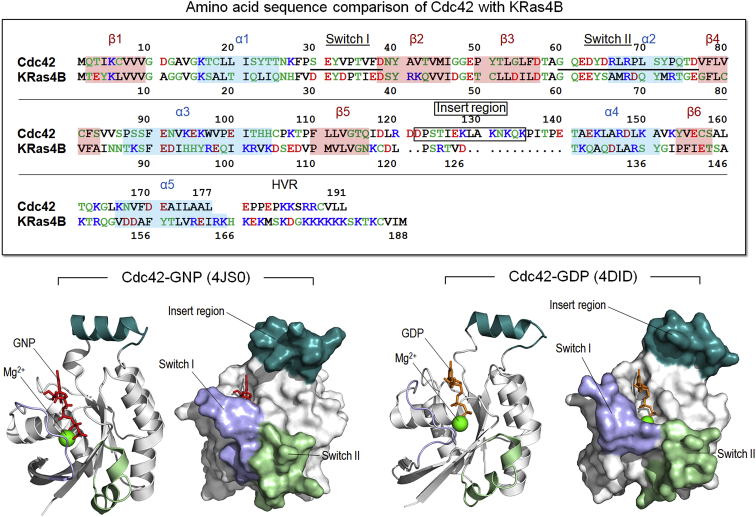
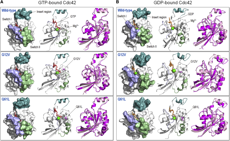
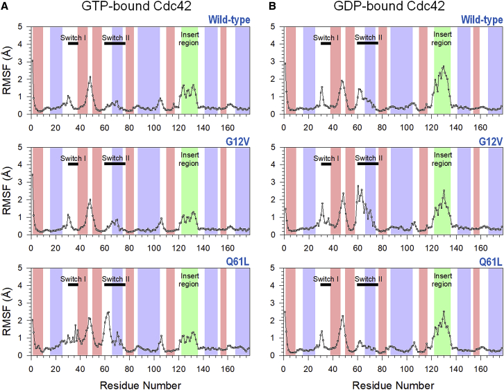
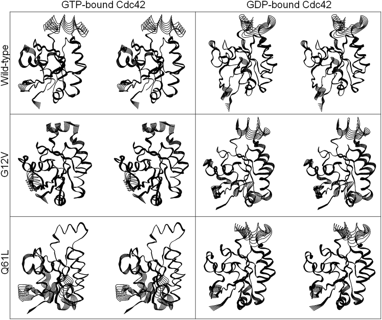
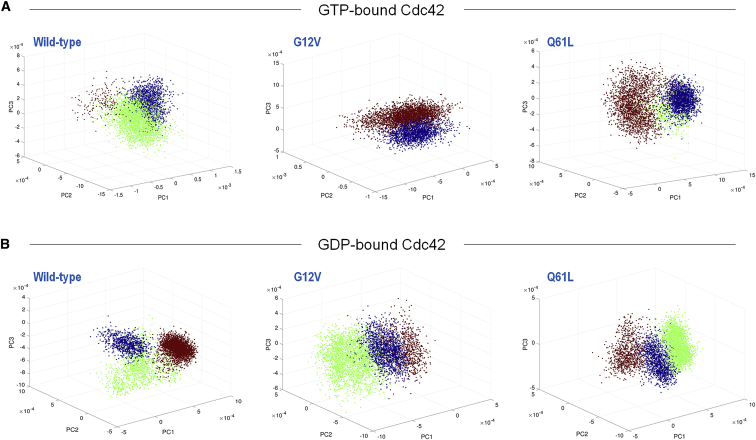
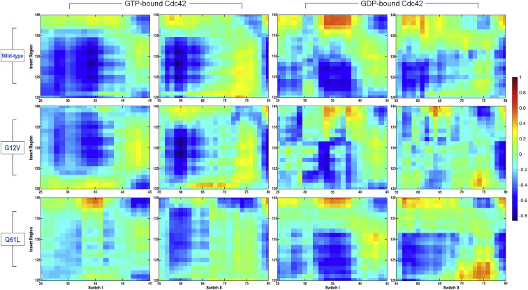
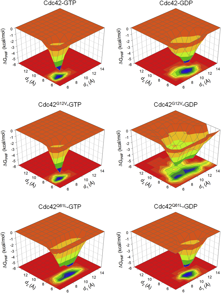
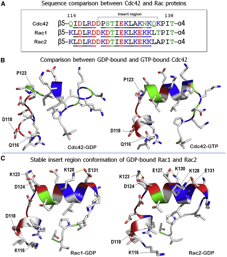
Cell division control protein 42 homolog (Cdc42) protein, a Ras superfamily GTPase, regulates cellular activities, including cancer progression. Using all-atom molecular dynamics (MD) simulations and essential dynamic analysis, we investigated the structure and dynamics of the catalytic domains of GDP-bound (inactive) and GTP-bound (active) Cdc42 in solution. We discovered substantial differences in the dynamics of the inactive and active forms, particularly in the "insert region" (residues 122-135), which plays a role in Cdc42 activation and binding to effectors. The insert region has larger conformational flexibility in the GDP-bound Cdc42 than in the GTP-bound Cdc42. The G2 loop and switch I at the effector lobe of the catalytic domain exhibit large conformational changes in both the GDP- and the GTP-bound systems, but in the GTP-bound Cdc42, the switch I interactions with GTP are retained. Oncogenic mutations were identified in the Ras superfamily. In Cdc42, the G12V and Q61L mutations decrease the GTPase activity. We simulated these mutations in both GDP- and GTP-bound Cdc42. Although the overall structural organization is quite similar between the wild type and the mutants, there are small differences in the conformational dynamics, especially in the two switch regions. Taken together, the G12V and Q61L mutations may play a role similar to their K-Ras counterparts in nucleotide binding and activation. The conformational differences, which are mainly in the insert region and, to a lesser extent, in the switch regions flanking the nucleotide binding site, can shed light on binding and activation. We propose that the differences are due to a network of hydrogen bonds that gets disrupted when Cdc42 is bound to GDP, a disruption that does not exist in other Rho GTPases. The differences in the dynamics between the two Cdc42 states suggest that the inactive conformation has reduced ability to bind to effectors.
Copyright © 2020 Biophysical Society. All rights reserved.
Figures
Similar articles
-
Molecular dynamics simulations reveal the activation mechanism of mutations G12V and Q61L of Cdc42.Proteins. 2022 Jul;90(7):1376-1389. doi: 10.1002/prot.26320. Epub 2022 Feb 21. Proteins. 2022. PMID: 35152498
-
Effector proteins exert an important influence on the signaling-active state of the small GTPase Cdc42.J Biol Chem. 2008 May 16;283(20):14153-64. doi: 10.1074/jbc.M706271200. Epub 2008 Mar 18. J Biol Chem. 2008. PMID: 18348980 Free PMC article.
-
Intrinsic GTP hydrolysis is observed for a switch 1 variant of Cdc42 in the presence of a specific GTPase inhibitor.Small GTPases. 2016;7(1):1-11. doi: 10.1080/21541248.2015.1123797. Epub 2016 Feb 1. Small GTPases. 2016. PMID: 26828437 Free PMC article.
-
Regulation of phosphorylation pathways by p21 GTPases. The p21 Ras-related Rho subfamily and its role in phosphorylation signalling pathways.Eur J Biochem. 1996 Dec 1;242(2):171-85. doi: 10.1111/j.1432-1033.1996.0171r.x. Eur J Biochem. 1996. PMID: 8973630 Review.
-
Targeting Cdc42 in cancer.Expert Opin Ther Targets. 2013 Nov;17(11):1263-73. doi: 10.1517/14728222.2013.828037. Epub 2013 Aug 19. Expert Opin Ther Targets. 2013. PMID: 23957315 Free PMC article. Review.
Cited by
-
Computational Investigation of Selected Spike Protein Mutations in SARS-CoV-2: Delta, Omicron, and Some Circulating Subvariants.Pathogens. 2023 Dec 21;13(1):10. doi: 10.3390/pathogens13010010. Pathogens. 2023. PMID: 38276156 Free PMC article.
-
Ras, RhoA, and vascular pharmacology in neurodevelopment and aging.Neurochem Int. 2024 Dec;181:105883. doi: 10.1016/j.neuint.2024.105883. Epub 2024 Oct 18. Neurochem Int. 2024. PMID: 39427854 Review.
-
Tumor-derived RHOA mutants interact with effectors in the GDP-bound state.Nat Commun. 2024 Aug 21;15(1):7176. doi: 10.1038/s41467-024-51445-z. Nat Commun. 2024. PMID: 39169042 Free PMC article.
-
A Natural Small Molecule Mitigates Kidney Fibrosis by Targeting Cdc42-mediated GSK-3β/β-catenin Signaling.Adv Sci (Weinh). 2024 Apr;11(13):e2307850. doi: 10.1002/advs.202307850. Epub 2024 Jan 19. Adv Sci (Weinh). 2024. PMID: 38240457 Free PMC article.
-
CDC42-IQGAP Interactions Scrutinized: New Insights into the Binding Properties of the GAP-Related Domain.Int J Mol Sci. 2022 Aug 9;23(16):8842. doi: 10.3390/ijms23168842. Int J Mol Sci. 2022. PMID: 36012107 Free PMC article.
References
Publication types
MeSH terms
Substances
Grants and funding
LinkOut - more resources
Full Text Sources
Other Literature Sources
Miscellaneous
