. 2020 Dec 17;21(24):9611.
doi: 10.3390/ijms21249611.

Vasa Vasorum Lumen Narrowing in Brain Vascular Hyalinosis in Systemic Hypertension Patients Who Died of Ischemic Stroke

Affiliations

Vasa Vasorum Lumen Narrowing in Brain Vascular Hyalinosis in Systemic Hypertension Patients Who Died of Ischemic Stroke

Sergiy G Gychka et al. Int J Mol Sci. .

Abstract

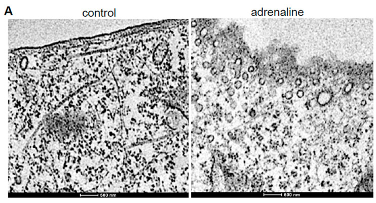
Ischemic stroke is a major cause of death among patients with systemic hypertension. The narrowing of the lumen of the brain vasculature contributes to the increased incidence of stroke. While hyalinosis represents the major pathological lesions contributing to vascular lumen narrowing and stroke, the pathogenic mechanism of brain vascular hyalinosis has not been well characterized. Thus, the present study examined the postmortem brain vasculature of human patients who died of ischemic stroke due to systemic hypertension. Hematoxylin and eosin staining and immunohistochemistry showed the occurrence of brain vascular hyalinosis with infiltrated plasma proteins along with the narrowing of the vasa vasorum and oxidative stress. Transmission electron microscopy revealed endothelial cell bulge protrusion into the vasa vasorum lumen and the occurrence of endocytosis in the vasa vasorum endothelium. The treatment of cultured microvascular endothelial cells with adrenaline also promoted the formation of the bulge as well as endocytic vesicles. The siRNA knockdown of sortin nexin-9 (a mediator of clathrin-mediated endocytosis) inhibited adrenaline-induced endothelial cell bulge formation. Adrenaline promoted protein-protein interactions between sortin nexin-9 and neural Wiskott-Aldrich syndrome protein (a regulator of actin polymerization). Spontaneously hypertensive stroke-prone rats also exhibited lesions indicative of brain vascular hyalinosis, the endothelial cell protrusion into the lumen of the vasa vasorum, and endocytosis in vasa vasorum endothelial cells. We propose that endocytosis-dependent endothelial cell bulge protrusion narrows the vasa vasorum, resulting in ischemic oxidative damage to cerebral vessels, the formation of hyalinosis, the occurrence of ischemic stroke, and death in systemic hypertension patients.

Keywords: brain; hyalinosis; ischemic stroke; oxidative stress; vasa vasorum; vascular.


Conflict of interest statement

The authors declare no conflict of interest. The funders had no role in the design of the study; in the collection, analyses, or interpretation of data; in the writing of the manuscript, or in the decision to publish the results.

Figures

Figure 1
Figure 1
Vascular hyalinosis in systemic hypertension patients who died of ischemic stroke. (A) Representative Zerbino–Lukasevich staining of patients with systemic hypertension who died of ischemic stroke. The left panel shows the infiltration of plasma fibrin into the vessel wall. The right panel (magnified, ×1000) shows the fibrin in the blood, at the vessel surface, and in the vessel wall. (B) Representative immunohistochemistry results using the ApoE antibody in patients who died of ischemic stroke without hypertension or with hypertension. Positive ApoE stains in the intima, media and adventitia layer were found only in the with hypertension group. Magnifications, ×400. Scale bars, 50 μm. Representative images of N = 8 patients per group.
Figure 2
Figure 2
H&E Staining of the vasa vasorum in the brain of patients who died of ischemic stroke. (A) Ischemic stroke without hypertension and with hypertension. Representative H&E staining images of n = 28 patients per group. Magnifications, ×400. Scale bar, 20 μm. (B) Quantification of vessel wall thickness. (C) Quantification of the cerebral vascular lumen area index calculated by dividing the internal surface area by the external surface area. n = 8 for the without hypertension group and n = 6 for the with hypertension group. (D) Quantification of vasa vasorum wall thickness. (E) Quantification of the vasa vasorum lumen area index calculated by dividing the internal surface area by the external surface area. n = 8. Mann–Whitney U test indicated that the values are significantly different at p < 0.05.
Figure 3
Figure 3
Immunohistochemistry for MDA. Representative immunohistochemistry results using the MDA antibody to assess the levels of oxidative stress in the brain of patients who died of ischemic stroke without hypertension or with hypertension. In patients with hypertension, the intense MDA expression is visible in the cerebral vessel walls (arrows) and the surrounding neuronal tissues. Magnifications ×400. Scale bars, 50 μm. Representative images of n = 8 patients per group.
Figure 4
Figure 4
TEM images of the vasa vasorum of the hyalinosis lesions of the brain vessels of systemic hypertension patients. Brain tissues from systemic hypertension patients who underwent neurosurgery to remove the hematoma to treat hemorrhagic stroke were analyzed by TEM. (A) Arrows indicate the presence of the endothelial cell bulge. Magnification, ×5600. (B) Arrows indicate the presence of endocytic vesicles. Magnification, ×22,000. Representative images of n = 6 patients.
Figure 5
Figure 5
Studies of cultured human microvascular endothelial cells. (A) TEM images of the control cells and cells exhibiting the adrenaline-induced formation of endocytic vesicles. Cells were treated with adrenaline at 10 μM for 4 h. Magnifications, ×13,000. (B) siRNA knockdown of SNX9 inhibits the adrenaline-induced bulge formation. Cells were treated with siRNA for 2 days and then with adrenaline at 10 μM for 4 h. Cells were observed under light microscopy. Arrows indicate the bulge formation. Magnifications, ×400. (C) Immunofluorescence staining of actin in untreated control and cells treated with adrenaline (10 μM; 4 h). The arrow indicates the reorganization of actin filaments. Magnifications, ×1000. (D) Adrenaline promotes SNX9–N-WASp interactions. Cells were treated with 10 μM adrenaline for 0, 1 or 2 h. Cell lysates were subjected to immunoprecipitation (IP) with mouse SNX9 IgG followed by Western blotting (WB) with rabbit N-WASp IgG.
Figure 5
Figure 5
Studies of cultured human microvascular endothelial cells. (A) TEM images of the control cells and cells exhibiting the adrenaline-induced formation of endocytic vesicles. Cells were treated with adrenaline at 10 μM for 4 h. Magnifications, ×13,000. (B) siRNA knockdown of SNX9 inhibits the adrenaline-induced bulge formation. Cells were treated with siRNA for 2 days and then with adrenaline at 10 μM for 4 h. Cells were observed under light microscopy. Arrows indicate the bulge formation. Magnifications, ×400. (C) Immunofluorescence staining of actin in untreated control and cells treated with adrenaline (10 μM; 4 h). The arrow indicates the reorganization of actin filaments. Magnifications, ×1000. (D) Adrenaline promotes SNX9–N-WASp interactions. Cells were treated with 10 μM adrenaline for 0, 1 or 2 h. Cell lysates were subjected to immunoprecipitation (IP) with mouse SNX9 IgG followed by Western blotting (WB) with rabbit N-WASp IgG.
Figure 6
Figure 6
Brain vascular hyalinosis in SHRSP rats. (A) H&E staining images of the brain vessels of Wistar–Kyoto (WKY) control rats and SHRSP rats at 8 weeks of age. Magnifications, ×400. (B) TEM image of the endothelial cell bulge protrusion into the lumen of the vasa vasorum in SHRSP rats. Magnification, ×14,500. (C) TEM image of endocytic vesicles in the endothelium of the vasa vasorum of SHRSP rats. Magnification, ×18,000. Representative images of n = 4 rats per group.
Figure 7
Figure 7
Scheme depicting the proposed mechanism of the endothelial cell (EC) bulge protrusion into the vasa vasorum lumen. In systemic hypertension patients, adrenaline promotes the endocytosis-dependent SNX9–N-WASp interaction that activates the actin polymerization and the EC bulge protrusion into the lumen of the vasa vasorum, resulting in narrowing the lumen of the vasa vasorum.

Similar articles

Cited by

References

    1. Benjamin E.J., Muntner P., Alonso A., Bittencourt M.S., Callaway C.W., Carson A.P., Chamberlain A.M., Chang A.R., Cheng S., Das S.R., et al. Heart disease and stroke Statistics-2019 update: A report from the American Heart Association. Circulation. 2019;139:e56–e528. doi: 10.1161/CIR.0000000000000659. - DOI - PubMed
    1. Heuschmann P.U., Wiedmann S., Wellwood I., Rudd A., Di Carlo A., Bejot Y., Ryglewicz D., Rastenyte D., Wolfe C.D., European Registers of Stroke Three-month stroke outcome: The European Registers of Stroke (EROS) investigators. Neurology. 2011;76:159–165. doi: 10.1212/WNL.0b013e318206ca1e. - DOI - PubMed
    1. Filipets O.O., Pashkovsky V.M. Stroke burden in Ukraine: Analysis of the official stroke statistics and overview of population-based epidemiological studies. Clin. Exp. Pathol. 2014;13:189–193.
    1. Ovbiagele B., Goldstein L.B., Higashida R.T., Howard V.J., Johnston S.C., Khavjou O.A., Lackland D.T., Lichtman J.H., Mohl S., Sacco R.L., et al. Forecasting the future of stroke in the United States. Stroke. 2013;44:2361–2375. doi: 10.1161/STR.0b013e31829734f2. - DOI - PubMed
    1. Kulesh S.D., Filina N.A., Frantava N.M., Zhytko N.L., Kastsinevich T.M., Kliatskova L.A., Shumskas M.S., Hilz M.J., Schwab S., Kolominsky-Rabas P.L. Incidence and case-fatality of stroke on the East border of the European union: The Grodno Stroke Study. Stroke. 2010;41:2726–2730. doi: 10.1161/STROKEAHA.110.596916. - DOI - PubMed

MeSH terms

Supplementary concepts