. 2020 Dec 17;21(24):9619.
doi: 10.3390/ijms21249619.

Effects of Fe2+/Fe3+ Binding to Human Frataxin and Its D122Y Variant, as Revealed by Site-Directed Spin Labeling (SDSL) EPR Complemented by Fluorescence and Circular Dichroism Spectroscopies

Affiliations

Effects of Fe2+/Fe3+ Binding to Human Frataxin and Its D122Y Variant, as Revealed by Site-Directed Spin Labeling (SDSL) EPR Complemented by Fluorescence and Circular Dichroism Spectroscopies

Davide Doni et al. Int J Mol Sci. .

Abstract

Frataxin is a highly conserved protein whose deficiency results in the neurodegenerative disease Friederich's ataxia. Frataxin's actual physiological function has been debated for a long time without reaching a general agreement; however, it is commonly accepted that the protein is involved in the biosynthetic iron-sulphur cluster (ISC) machinery, and several authors have pointed out that it also participates in iron homeostasis. In this work, we use site-directed spin labeling coupled to electron paramagnetic resonance (SDSL EPR) to add new information on the effects of ferric and ferrous iron binding on the properties of human frataxin in vitro. Using SDSL EPR and relating the results to fluorescence experiments commonly performed to study iron binding to FXN, we produced evidence that ferric iron causes reversible aggregation without preferred interfaces in a concentration-dependent fashion, starting at relatively low concentrations (micromolar range), whereas ferrous iron binds without inducing aggregation. Moreover, our experiments show that the ferrous binding does not lead to changes of protein conformation. The data reported in this study reveal that the currently reported binding stoichiometries should be taken with caution. The use of a spin label resistant to reduction, as well as the comparison of the binding effect of Fe2+ in wild type and in the pathological D122Y variant of frataxin, allowed us to characterize the Fe2+ binding properties of different protein sites and highlight the effect of the D122Y substitution on the surrounding residues. We suggest that both Fe2+ and Fe3+ might play a relevant role in the context of the proposed FXN physiological functions.

Keywords: CD; EPR; Fe-S cluster assembly machinery; fluorescence; frataxin; iron.


Conflict of interest statement

The authors declare no conflict of interest.

Figures

Scheme 1
Scheme 1
The MTSSL and the M-TETPO nitroxide spin labels.
Figure 1
Figure 1
Structure of human FXN (PDB.ID 1EKG) from different angles, the N-terminus is in dark grey, the C-terminus is white. The Glu and Asp residues are colored dark and light orange, respectively. The three native Trp residues are colored bright green and shown in ball and stick representation. The spin labeling sites—A99 (blue), A114 (yellow), T133 (green), H183 (pink), A193 (red)—are shown in stick representation. The D122 residue in light orange, the site of the pathological D122Y mutation, is shown in ball and stick representation.
Figure 2
Figure 2
EPR spectra of FXN mutants at increasing protein: Fe3+ molar ratios from 1:0 to 1:50; protein concentration 10 µM, MTSSL label. The colors are the same of the positions highlighted in Figure 1: (A) A114C—brown; (B) A114C/D122Y—brown; (C) T133C—green; (D) H183C—pink; (E) A193C—red. Central and Right panels: EPR spectra (black) and simulations (color) of FXN mutants at protein: Fe3+ molar ratios 1:0 (central) and 1:50 (right).
Figure 3
Figure 3
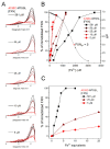
(A) From top to bottom, the left shoulder of the EPR spectrum of the A193C mutant labeled with MTSSL at progressively lower protein concentrations, the lighter the color the higher the FXN: Fe3+ ratio. (B) The percentage of immobilized protein (left axis, big dots) or the pH (right axis, small dots) vs. the nominal ferric iron concentration for different protein concentrations. (C) The percentage of immobilized protein vs. the FXN: Fe3+ molar ratio for three protein concentrations.
Figure 4
Figure 4
EPR spectra of different FXN mutants at increasing protein: Fe2+ molar ratios from 1:0 to 1:50; protein concentration 50 µM, M-TETPO label. (A) A99C—blue; (B) T133C—green; (C) A114C—brown; (D) H183C—pink; (E) A114C-D122Y brown; (F) A193C—red.
Figure 5
Figure 5
Tryptophan fluorescence spectra of WT FXN and D122Y variant at increasing amounts of Fe3+. Protein concentration 1.4 µM, 288 K, λexc = 280 nm, λmax = 332 nm. (A,B) Fluorescence of FXN WT and D122Y, respectively; (C,D) fluorescence quenching by Fe3+ calculated at λmax for FXN WT and D122Y, respectively; in the inset, a zoom of the sub-stoichiometric region.
Figure 6
Figure 6
Tryptophan fluorescence spectra of WT FXN and D122Y variant at increasing amounts of Fe2+. Protein concentration 1.4 µM, 288 K, λexc = 280 nm, λmax = 332 nm. (A,B) Fluorescence of FXN WT and D122Y, respectively; (C,D) fluorescence quenching by Fe2+ calculated at λmax for FXN WT and D122Y, respectively; in the inset, a zoom of the sub-stoichiometric region.
Figure 7
Figure 7
CD spectra of FXN of WT FXN and D122Y variant with either Fe3+ or Fe2+. Protein concentration 50 µM. (A) WT FXN with increasing amounts of Fe3+; (B) D122Y FXN with increasing amounts of Fe3+; (C) WT FXN with Fe2+; (D) D122Y FXN with Fe2+.
Scheme 2
Scheme 2
Synthetic way to prepare TETPO from alcohol 1. (a) MsCl, Et3N, DCM 0 °C to RT, 2 h; (b) protected maleimide, K2CO3, DMF; (c) reflux, toluene.

Similar articles

Cited by

References

    1. Campuzano V., Montermini L., Molto M.D., Pianese L., Cossee M., Cavalcanti F., Monros E., Rodius F., Duclos F., Monticelli A., et al. Friedreich’s Ataxia: Autosomal Recessive Disease Caused by an Intronic GAA Triplet Repeat Expansion. Science. 1996;271:1423–1427. doi: 10.1126/science.271.5254.1423. - DOI - PubMed
    1. Galea C.A., Huq A., Lockhart P.J., Tai G., Corben L.A., Yiu E.M., Gurrin L.C., Lynch D.R., Gelbard S., Durr A., et al. Compound Heterozygous FXN Mutations and Clinical Outcome in Friedreich Ataxia. Ann. Neurol. 2016;79:485–495. doi: 10.1002/ana.24595. - DOI - PubMed
    1. De Castro M., García-Planells J., Monrós E., Cañizares J., Vázquez-Manrique R., Vílchez J.J., Urtasun M., Lucas M., Navarro G., Izquierdo G., et al. Genotype and Phenotype Analysis of Friedreich’s Ataxia Compound Heterozygous Patients. Hum. Genet. 2000;106:86–92. doi: 10.1007/s004399900201. - DOI - PubMed
    1. Gellera C., Castellotti B., Mariotti C., Mineri R., Seveso V., DiDonato S., Taroni F. Frataxin Gene Point Mutations in Italian Friedreich Ataxia Patients. Neurogenetics. 2007;8:289–299. doi: 10.1007/s10048-007-0101-5. - DOI - PubMed
    1. Chiabrando D., Bertino F., Tolosano E. Hereditary Ataxia: A Focus on Heme Metabolism and Fe-S Cluster Biogenesis. Int. J. Mol. Sci. 2020;21:3760. doi: 10.3390/ijms21113760. - DOI - PMC - PubMed