. 2020 Dec 17;21(24):9631.
doi: 10.3390/ijms21249631.

Suppression of Pax3-MITF-M Axis Protects from UVB-Induced Skin Pigmentation by Tetrahydroquinoline Carboxamide

Affiliations

Suppression of Pax3-MITF-M Axis Protects from UVB-Induced Skin Pigmentation by Tetrahydroquinoline Carboxamide

Yong-Pyo Choi et al. Int J Mol Sci. .

Abstract

Paired box gene 3 (Pax3) and cAMP responsive element-binding protein (CREB) directly interact with the cis-acting elements on the promoter of microphthalmia-associated transcription factor isoform M (MITF-M) for transcriptional activation in the melanogenic process. Tyrosinase (Tyro) is a target gene of MITF-M, and functions as a key enzyme in melanin biosynthesis. Tetrahydroquinoline carboxamide (THQC) was previously screened as an antimelanogenic candidate. In the current study, we evaluated the antimelanogenic activity of THQC in vivo and elucidated a possible mechanism. Topical treatment with THQC mitigated ultraviolet B (UVB)-induced skin pigmentation in guinea pig with decreased messenger RNA (mRNA) and protein levels of melanogenic genes such as MITF-M and Tyro. Moreover, THQC inhibited cAMP-induced melanin production in α-melanocyte-stimulating hormone (α-MSH)- or histamine-activated B16-F0 cells, in which it suppressed the expression of the MITF-M gene at the promoter level. As a mechanism, THQC normalized the protein levels of Pax3, a transcriptional activator of the MITF-M gene, in UVB-exposed and pigmented skin, as well as in α-MSH-activated B16-F0 culture. However, THQC did not affect UVB- or α-MSH-induced phosphorylation (activation) of CREB. The results suggest that suppression of the Pax3-MITF-M axis might be a potential strategy in the treatment of skin pigmentary disorders that are at high risk under UVB radiation.

Keywords: guinea pig skin; melanin pigmentation; microphthalmia-associated transcription factor isoform M; paired box gene 3; tetrahydroquinoline carboxamide; tyrosinase.


Conflict of interest statement

The authors declare no conflict of interest.

Figures
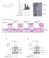
Figure 1
Figure 1
Effect of tetrahydroquinoline carboxamide (THQC) on skin pigmentation in guinea pig. (A) Chemical structure of THQC. (BE) Dorsal skin of guinea pig was irradiated with ultraviolet B (UVB) and treated topically with THQC according to the protocol in Figure S1 (Supplementary Materials). (B) The melanin index was measured in UVB-exposed area of skin and is represented as relative fold. A photograph of pigmented skin is also presented. The vehicle was a mixture of propylene glycol, ethanol, and water (5:3:2). (C) Skin tissue was sectioned and then reacted with Fontana–Masson silver nitrate to stain melanin granule as black. (D) Protein extract from skin tissue was resolved on SDS acrylamide gel by electrophoresis and subjected to Western blot analysis (WB) with anti-tyrosinase (Tyro), anti-microphthalmia-associated transcription factor (MITF), or anti-glyceraldehyde 3-phosphate dehydrogenase (GAPDH) antibody. (E) Total RNA from skin tissue was subjected to semi-quantitative RT-PCR analysis of Tyro or MITF-M with the internal control β-actin and resolved on agarose gel by electrophoresis. * p < 0.05 vs. UVB alone.
Figure 2
Figure 2
Effect of THQC on melanin production in B16-F0 culture. B16-F0 cells were stimulated with α-melanocyte-stimulating hormone (α-MSH) (A), histamine (B), or dibutyryl (db)-cAMP (C) for 72 h in the presence of THQC. Melanin pigment was quantified by measuring absorbance value at 405 nm and is represented as relative fold. * p < 0.05 vs. α-MSH, histamine, or db-cAMP alone.
Figure 3
Figure 3
Effect of THQC on Tyro expression. B16-F0 cells were stimulated with α-MSH for 48 h (A,B) or 20 h (C) in the presence of THQC. (A) Cell extract as the enzyme source of Tyro was reacted with l-dopa as a substrate in sodium phosphate buffer, and immediately measured the increase in absorbance value at 475 nm per min. Tyro activity is represented as the initial velocity of dopa oxidation (nmol/min). (B) Protein extract was resolved on SDS acrylamide gel by electrophoresis and subjected to Western blot analysis (WB) with anti-Tyro or anti-GAPDH antibody. (C) Total RNA was subjected to semi-quantitative RT-PCR analysis of Tyro with the internal control β-actin and resolved on agarose gene by electrophoresis. (D) B16-F0 cells harboring the Tyro (−2236/+59)-Luc reporter in combination with the Renilla control vector were stimulated with α-MSH for 18 h in the presence of THQC. Firefly luciferase activity, reporting the promoter activity of Tyro gene, was normalized to the Renilla activity and is represented as relative fold. (E) Tyro protein was treated with THQC and reacted with l--dopa as a substrate in cell-free reactions. Tyro activity is represented as the initial velocity of dopa oxidation (nmol/min). * p < 0.05 vs. α-MSH alone (AD) or Tyro protein alone (E).
Figure 4
Figure 4
Effect of THQC on MITF-M expression. After pretreating with THQC for 2 h, B16-F0 cells were stimulated with α-MSH for 4 h (A) or 2 h (B) in the presence of THQC. (A) Western blot analysis (WB) of MITF with the internal control GAPDH. (B) Semi-quantitative RT-PCR analysis of MITF-M with the internal control β-actin. (C) After transfection with the MITF-M (−2200/+95)-Luc reporter in combination with the Renilla control vector, B16-F0 cells were stimulated with α-MSH for 18 h in the presence of THQC. The cell extract was subjected to a dual luciferase assay. Firefly luciferase activity, reporting the promoter activity of the MITF-M gene, is represented as relative fold after normalizing to Renilla activity. * p < 0.05 vs. α-MSH alone.
Figure 5
Figure 5
Effect of THQC on transcription factors regulating the MITF-M promoter. Transcription factors paired box gene 3 (Pax3), cAMP responsive element-binding protein (CREB), and β-catenin directly interact with the MITF-M promoter for transcriptional activation. The dorsal skin of guinea pig was irradiated with UVB and treated topically with THQC according to the protocol in Figure S1 (Supplementary Materials) (A,C). B16-F0 cells were pretreated with THQC for 2 h and stimulated with α-MSH for 2 h (B) or 15 min (D) in the presence of THQC. (A) Protein extract from skin tissue was resolved on SDS acrylamide gel by electrophoresis and subjected to Western blot analysis (WB) with anti-Pax3 or anti-GAPDH antibody. (B) Protein extract from B16-F0 culture was subjected to WB with anti-Pax3 or anti-GAPDH antibody. (C) Protein extract from skin tissue was subjected to WB with anti-phosphorylated (p)-CREB or anti-CREB antibody. (D) Protein extract from B16-F0 culture was subjected to WB with anti-p-CREB, anti-CREB, anti-p-β-catenin, or anti-β-catenin antibody. * p < 0.05 vs. UVB alone (A) or α-MSH alone (B,D).
Figure 6
Figure 6
A proposed mechanism of THQC on antimelanogenic activity. Topical treatment with THQC mitigated facultative pigmentation in UVB-irradiated skin via blocking the Pax3–MITF-M axis.

Similar articles

Cited by

References

    1. Yamaguchi Y., Hearing V.J. Physiological factors that regulate skin pigmentation. Biofactors. 2009;35:193–199. doi: 10.1002/biof.29. - DOI - PMC - PubMed
    1. Miyamura Y., Coelho S.G., Wolber R., Miller S.A., Wakamatsu K., Zmudzka B.Z., Ito S., Smuda C., Passeron T., Choi W., et al. Regulation of human skin pigmentation and responses to ultraviolet radiation. Pigment Cell Res. 2007;20:2–13. doi: 10.1111/j.1600-0749.2006.00358.x. - DOI - PubMed
    1. Slominski A.T., Zmijewski M.A., Plonka P.M., Szaflarski J.P., Paus R. How UV light touches the brain and endocrine system through skin, and why. Endocrinology. 2018;159:1992–2007. doi: 10.1210/en.2017-03230. - DOI - PMC - PubMed
    1. Yuan X.H., Jin Z.H. Paracrine regulation of melanogenesis. Br. J. Dermatol. 2018;178:632–639. doi: 10.1111/bjd.15651. - DOI - PubMed
    1. Slominski A., Wortsman J., Luger T., Paus R., Solomon S. Corticotropin releasing hormone and proopiomelanocortin involvement in the cutaneous response to stress. Physiol. Rev. 2000;80:979–1020. doi: 10.1152/physrev.2000.80.3.979. - DOI - PubMed

MeSH terms

Substances