Review
. 2020 Dec 17;10(12):355.
doi: 10.3390/life10120355.

Structural Determinants and Their Role in Cyanobacterial Morphogenesis

Affiliations
Review

Structural Determinants and Their Role in Cyanobacterial Morphogenesis

Benjamin L Springstein et al. Life (Basel). .

Abstract

Cells have to erect and sustain an organized and dynamically adaptable structure for an efficient mode of operation that allows drastic morphological changes during cell growth and cell division. These manifold tasks are complied by the so-called cytoskeleton and its associated proteins. In bacteria, FtsZ and MreB, the bacterial homologs to tubulin and actin, respectively, as well as coiled-coil-rich proteins of intermediate filament (IF)-like function to fulfil these tasks. Despite generally being characterized as Gram-negative, cyanobacteria have a remarkably thick peptidoglycan layer and possess Gram-positive-specific cell division proteins such as SepF and DivIVA-like proteins, besides Gram-negative and cyanobacterial-specific cell division proteins like MinE, SepI, ZipN (Ftn2) and ZipS (Ftn6). The diversity of cellular morphologies and cell growth strategies in cyanobacteria could therefore be the result of additional unidentified structural determinants such as cytoskeletal proteins. In this article, we review the current advances in the understanding of the cyanobacterial cell shape, cell division and cell growth.

Keywords: FtsZ; IF proteins; MreB; cell division; cell shape; cyanobacteria; cytoskeleton; morphology.


Conflict of interest statement

The authors declare no conflict of interest. The funders had no role in the design of the study; in the collection, analyses, or interpretation of data; in the writing of the manuscript, or in the decision to publish the results.

Figures
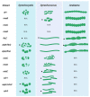
Figure 1
Figure 1
Cyanobacterial cell division and cell growth mutant phenotypes in Synechocystis, Synechococcus, and Anabaena. Stars indicate gene essentiality in the respective organism. Of note: while one gene can be essential in one cyanobacterial organism/morphotype, it does not necessarily mean it is essential in all other cyanobacteria. N/A indicates that no mutant phenotypes have been described. WT: wild type. Image created with BioRender.com. WT [19]; mreB [58,81,83,84]; mreC [58]; mreD [58]; ftsZ [86,87]; zipN/ftn2 [86,88,89]; zipS/ftn6 [86,89,90]; minC [88,91]; minD [88,91]; minE [88,91]; cdv1 [86]; sepF/cdv2 [86,90]; cdv3 [86,92].
Figure 2
Figure 2
Proposed model of the Anabaena divisome. Proteins in grey shades are inferred from a previous model described for Synechocystis [92]. PatA is assumed to negatively interfere with the linkage of FtsZ to the cytoplasmic membrane through the loss of interaction with its presumed cytoplasmic membrane anchors SepF and ZipN.
Figure 3
Figure 3
Interaction network of cell division/growth proteins in Synechocystis and Anabaena. Depiction of protein-protein interactions as identified by bacterial adenylate cyclase two hybrid assays and co-immunoprecipitation experiments. Interactions were identified in [87,88,90,92,132,172]. Black arrows indicate interactions solely found in one species so far, while purple arrows mark interactions found in both Synechocystis and Anabaena. Interactions only attributed to one species do not necessarily imply these interactions do not exist in the other but rather that these interactions were not yet tested for. Image created with BioRender.com.
Figure 4
Figure 4
Bacterial coiled-coil-rich proteins. Depiction of coiled-coil-rich-regions (purple rectangles) in (A) the human vimentin, (B) previously described bacterial CCRPs, and (C) recently identified cyanobacterial CCRPs. Coiled-coil-rich regions were predicted using the COILS algorithm [212] or were obtained from [9,197,204,211]. Orange rectangles indicate TPR repeats that are also identified as coiled-coils by the COILS algorithm. Vimentin [188]; TlpA [192]; Crescentin [9]; FilP [197], Syc2039, HmpFSyc, Slr7083, HmpFSyn, Fm7001, All4981 [204]; ZicK, ZacK [211].
Figure 5
Figure 5
Electron micrographs of Anabaena. (A) Thin section of Anabaena minutissima showing striated microtubules (indicated by arrows). Pb, polyhedral bodies; CG, cyanophycin granule. (B,C) Cryo-electron tomograms of Anabaena showing uncharacterized intracellular filaments. (B) 5 nm wide filaments, with repetitive units every 5.5 nm, bundled together in the cytoplasm (arrowheads) and spanning parts of the cell. (C) Cross-section of these bundles revealed a tight packing with 11 nm spacing between filament centres (arrow). IM, inner membrane; OM, outer membrane; PB, phycobilisomes; PG, peptidoglycan; TM, thylakoid membrane. Bars, 100 nm. Shown are 13.5 nm thick slices. Figure 5A is reprinted from “The fine structure of striated microtubules and sleeve bodies in several species of Anabaena” [215], vol. 57, 1976, by Thomas E. Jensen and Robert P. Ayala with permission from Elsevier.

Similar articles

Cited by

References

    1. Moseley J.B. An expanded view of the eukaryotic cytoskeleton. Mol. Biol. Cell. 2013;24:1615–1618. doi: 10.1091/mbc.e12-10-0732. - DOI - PMC - PubMed
    1. Alberts B., Johnson A., Lewis J., Morgan D., Raff M., Roberts K., Walter P. Molecular Biology of the Cell. 6th ed. Garland Science; New York, NY, USA: 2014.
    1. Huber F., Boire A., López M.P., Koenderink G.H. Cytoskeletal crosstalk: When three different personalities team up. Curr. Opin. Cell Biol. 2015;32:39–47. doi: 10.1016/j.ceb.2014.10.005. - DOI - PubMed
    1. Bi E., Lutkenhaus J. FtsZ ring structure associated with division in Escherichia coli. Nature. 1991;354:161–164. doi: 10.1038/354161a0. - DOI - PubMed
    1. De Boer P., Crossley R., Rothfield L. The essential bacterial cell-division protein FtsZ is a GTPase. Nature. 1992;359:254–256. doi: 10.1038/359254a0. - DOI - PubMed

LinkOut - more resources