. 2020 Dec 17;9(12):4084.
doi: 10.3390/jcm9124084.

Identification of Markers Predicting Clinical Course in Patients with IgG4-Related Ophthalmic Disease by Unbiased Clustering Analysis

Affiliations

Identification of Markers Predicting Clinical Course in Patients with IgG4-Related Ophthalmic Disease by Unbiased Clustering Analysis

Kinya Tsubota et al. J Clin Med. .

Abstract

Purpose: To describe the clinical features of patients with immunoglobulin G4 (IgG4)-related ophthalmic disease (IgG4-ROD) grouped by unbiased cluster analysis using peripheral blood test data and to find novel biomarkers for predicting clinical features.

Methods: One hundred and seven patients diagnosed with IgG4-ROD were divided into four groups by unsupervised hierarchical cluster analysis using peripheral blood test data. The clinical features of the four groups were compared and novel markers for prediction of clinical course were explored.

Results: Unbiased cluster analysis divided patients into four groups. Group B had a significantly higher frequency of extraocular muscle enlargement (p < 0.001). The frequency of patients with decreased best corrected visual acuity (BCVA) was significantly higher in group D (p = 0.002). Receiver operating characteristic (ROC) curves for the prediction of extraocular muscle enlargement and worsened BCVA using a panel consisting of important blood test data identified by machine learning yielded areas under the curve of 0.78 and 0.86, respectively. Clinical features were compared between patients divided into two groups by the cutoff serum IgE or IgG4 level obtained from ROC curves. Patients with serum IgE above 425 IU/mL had a higher frequency of extraocular muscle enlargement (25% versus 6%, p = 0.004). Patients with serum IgG4 above 712 mg/dL had a higher frequency of decreased BCVA (37% versus 5%, p ≤ 0.001).

Conclusion: Unsupervised hierarchical clustering analysis using routine blood test data differentiates four distinct clinical phenotypes of IgG4-ROD, which suggest differences in pathophysiologic mechanisms. High serum IgG4 is a potential predictor of worsened BCVA, and high serum IgE is a potential predictor of extraocular muscle enlargement in IgG4-ROD patients.

Keywords: IgG4-related ophthalmic disease; biomarker; decreased best corrected visual acuity; extraocular muscle enlargement; unbiased cluster analysis.


Conflict of interest statement

No conflicting relationship exists for any author.

Figures
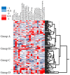
Figure 1
Figure 1
Patients are clustered into four groups by unsupervised hierarchical clustering analysis using the data of 20 peripheral blood tests. Group A, patients with low white blood cell (WBC) count; group B, patients with high serum immunoglobulin E (IgE); group C, patients with high Plt count; and group D, patients with high serum IgG4.
Figure 2
Figure 2
ROC curves for extraocular muscle enlargement, worsened best corrected visual acuity (BCVA), and lesion above the neck excluding LG and SG using significantly different laboratory findings obtained from comparison of laboratory findings in patients with and those without specific clinical features. (A). Receiver operating characteristic (ROC) curves for extraocular muscle enlargement using WBC count, Eo count, C-reactive protein (CRP), and IgE. Area under the ROC curve (AUC) of IgE is the largest among the four markers (0.71). Cutoff value of IgE is 425 IU/mL, with the highest odds ratio. (B). ROC curves for worsened BCVA using serum IgG4, β2MG, serum IgE, serum IgA, serum IgG, CRP, serum TP, and WBC count. AUC of IgG4 is the largest among the eight markers (0.80). Cutoff value of IgG4 is 713 mg/dL, with the highest odds ratio. (C). ROC curve for lesion above the neck excluding LG and SG using peripheral WBC count. AUC of WBC is 0.63. Cutoff value of WBC count is 6700 number/μL.
Figure 3
Figure 3
Machine learning analysis using random forest by Boruta multivariate analysis for the prediction of clinical features. Green box denotes a feature confirmed to be important; yellow box denotes a tentative attribute, red box denotes unimportant, and blue box denotes the shadow. (A). IgE, Eo count, and T-Bil (three green boxes) are more important for the prediction of extraocular muscle enlargement than other peripheral blood findings. (B). WBC, T-Bil, serum IgG, serum IgA, β2-MG, and serum IgG4 (six green boxes) are more important for the prediction of worsened BCVA than other peripheral blood findings. (C). All peripheral blood findings are not important for the prediction of lesion above the neck excluding LG and SG. °, * outlier.
Figure 4
Figure 4
ROC curves for extraocular muscle enlargement (A) and worsened BCVA (B) using peripheral blood tests identified by Boruta multivariate analysis shown in Figure 3. A. AUC is 0.78 by ROC analysis using three green boxes in Figure 3B (IgE, number of Eo, and T-BIL). B. AUC is 0.86 by ROC analysis using six green boxes in Figure 3B (WBC, T-BIL, serum IgG, serum IgA, β2-MG, and serum IgG4).

Similar articles

Cited by

References

    1. Hamano H., Kawa S., Horiuchi A., Unno H., Furuya N., Akamatsu T., Fukushima M., Nikaido T., Nakayama K., Usuda N. High serum IgG4 concentrations in patients with sclerosing pancreatitis. N. Engl. J. Med. 2001;344:732–738. doi: 10.1056/NEJM200103083441005. - DOI - PubMed
    1. Yamamoto M., Ohara M., Suzuki C., Naishiro Y., Yamamoto H., Takahashi H., Imai K. Elevated IgG4 concentrations in serum of patients with Mikulicz’s disease. Scand. J. Rheumatol. 2004;33:432–433. doi: 10.1080/03009740410006439. - DOI - PubMed
    1. Stone J.H., Khosroshahi A., Deshpande V., Chan J.K., Heathcote J.G., Aalberse R., Azumi A., Bloch D.B., Brugge W.R., Carruthers M.N. Recommendations for the nomenclature of IgG4-related disease and its individual organ system manifestations. Arthritis Rheum. 2012;64:3061–3067. doi: 10.1002/art.34593. - DOI - PMC - PubMed
    1. Umehara H., Okazaki K., Masaki Y., Kawano M., Yamamoto M., Saeki T., Matsui S., Yoshino T., Nakamura S., Kawa S. Comprehensive diagnostic criteria for IgG4-related disease (IgG4-RD) Mod. Rheumatol. 2012;22:21–30. doi: 10.3109/s10165-011-0571-z. - DOI - PubMed
    1. Wallace Z.S., Zhang Y., Perugino C.A., Naden R., Choi H.K., Stone J.H., ACR/EULAR IgG4-RD Classification Criteria Committee Clinical phenotypes of IgG4-related disease: an analysis of two international cross-sectional cohorts. Ann. Rheum. Dis. 2019;78:406–412. doi: 10.1136/annrheumdis-2018-214603. - DOI - PMC - PubMed

LinkOut - more resources