. 2020 Dec 17;10(12):357.
doi: 10.3390/life10120357.

β-Catenin Regulates Cardiac Energy Metabolism in Sedentary and Trained Mice

Affiliations

β-Catenin Regulates Cardiac Energy Metabolism in Sedentary and Trained Mice

Volodymyr V Balatskyi et al. Life (Basel). .

Abstract

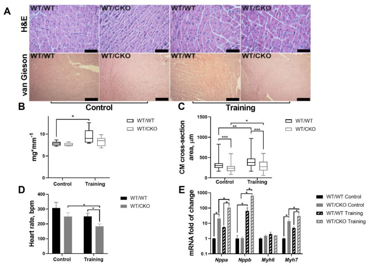
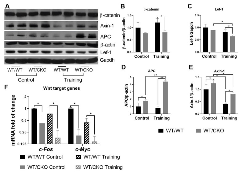
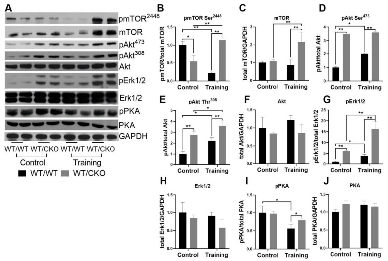
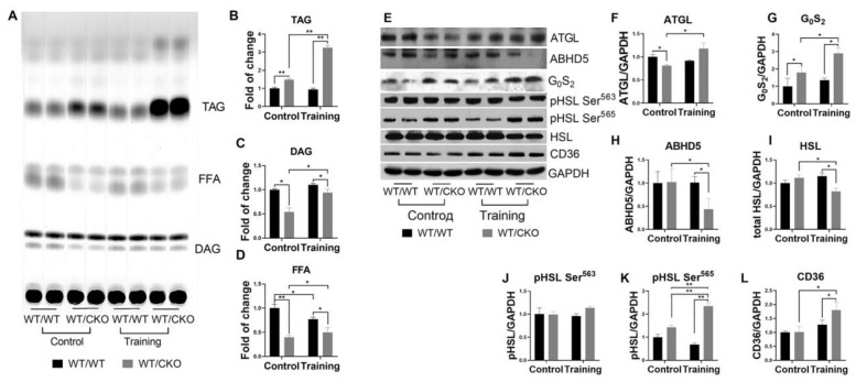
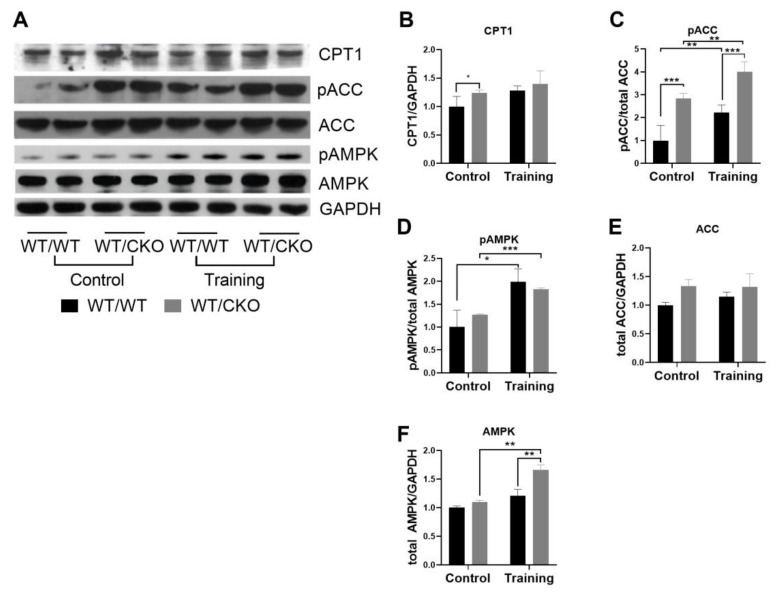
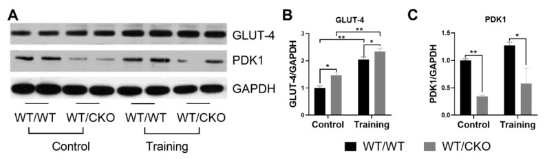
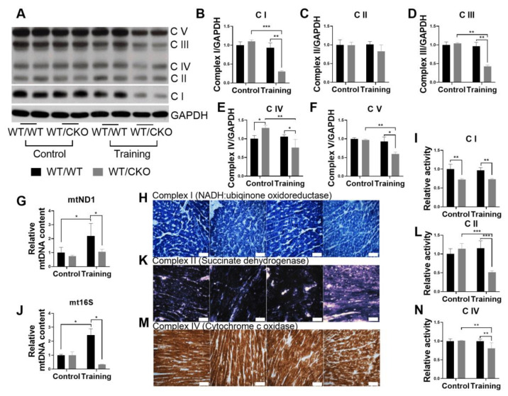
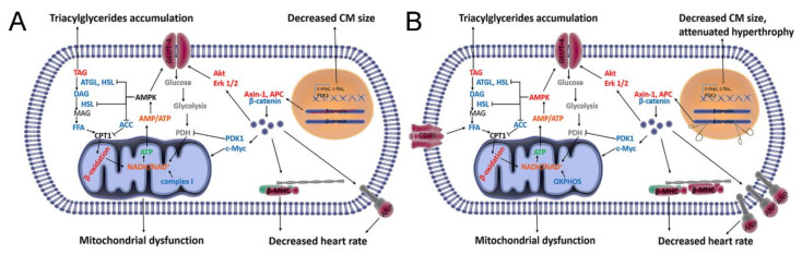
The role of canonical Wnt signaling in metabolic regulation and development of physiological cardiac hypertrophy remains largely unknown. To explore the function of β-catenin in the regulation of cardiac metabolism and physiological cardiac hypertrophy development, we used mice heterozygous for cardiac-specific β-catenin knockout that were subjected to a swimming training model. β-Catenin haploinsufficient mice subjected to endurance training displayed a decreased β-catenin transcriptional activity, attenuated cardiomyocytes hypertrophic growth, and enhanced activation of AMP-activated protein kinase (AMPK), phosphoinositide-3-kinase-Akt (Pi3K-Akt), and mitogen-activated protein kinase/extracellular signal-regulated kinases 1/2 (MAPK/Erk1/2) signaling pathways compared to trained wild type mice. We further observed an increased level of proteins involved in glucose aerobic metabolism and β-oxidation along with perturbed activity of mitochondrial oxidative phosphorylation complexes (OXPHOS) in trained β-catenin haploinsufficient mice. Taken together, Wnt/β-catenin signaling appears to govern metabolic regulatory programs, sustaining metabolic plasticity in adult hearts during the adaptation to endurance training.

Keywords: Wnt/β-catenin signaling; glucose metabolism; lipid metabolism; oxidative phosphorylation; training-induced heart hypertrophy; β-oxidation.


Conflict of interest statement

The authors declare no conflict of interest. The authors alone are responsible for the views expressed in this article and they do not necessarily represent the views, decisions, or policies of the institutions with which they are affiliated. The funders had no role in the design of the study; in the collection, analyses, or interpretation of data; in the writing of the manuscript, or in the decision to publish the results.

Figures

Figure 1
Figure 1
Heterozygous deletion of β-catenin does not affect adult heart morphology, but attenuates the hypertrophic response. (A) Top row— hematoxylin and eosin (H&E)-stained paraffin-embedded sections; 400× magnification, scale bar—50 μm. Bottom row—Picrofuchsin van Gieson-stained paraffin-embedded sections; 100× magnification, scale bar—200 μm for H&E and 100× for van Gieson; WT/WT—control; WT/CKO—heterozygous. (B) Heart weight/tibia length (HW/TL) ratio in trained and untrained mice. WT/WT control, n = 11, WT/WT training n = 11, WT/CKO control, n = 7, WT/CKO training n = 12. (C) Cardiomyocytes cross-section area in trained and untrained mice. Over 100 cells per heart from 3 hearts of each genotype were analyzed. (D) Electrophysiological monitoring of heart rate (beat/min) in trained and untrained mice. WT/WT control, n = 5, WT/WT training, n = 4, WT/CKO control, n = 4, WT/CKO training n = 4. (E) qPCR analysis of fetal gene expression in control and trained WT/WT and CKO/WT mice. n = 5. The data are expressed as the mean ± SD of arbitrary fold of change relative to control levels. * p < 0.05, ** p < 0.01, *** p < 0.005 (Kruskal–Wallis test followed by Dunn’s multiple-comparison post hoc test).
Figure 2
Figure 2
Heterozygous knockout of β-catenin leads to canonical Wnt signaling downregulation in the hearts of trained and untrained mice. (A) Western blot of total β-catenin, Lef-1, Axin-1, and adenomatous polyposis coli (APC) in left ventricle (LV) lysates from WT/WT and WT/CKO mice in sedentary conditions (Control) and after endurance training (Training). (B) Densitometry of total β-catenin normalized to total β-actin. (C) Densitometry of Lef-1 normalized to Gapdh. (D) Densitometry of Axin-1 normalized to β-actin. (E) Densitometry of APC normalized to β-actin. (F) qPCR analysis of β-catenin target gene expression. The data are expressed as the mean ± SD of arbitrary fold of change relative to control levels. n = 5/group. * p < 0.05, ** p < 0.01 (Kruskal–Wallis test followed by Dunn’s multiple-comparison post hoc test).
Figure 3
Figure 3
Heterozygous knockout of β-catenin affects hypertrophic signaling in the heart. (A) Western blot of pmTOR2448, mTOR, pAkt at Thr308, pAkt at Ser473, Akt, pErk1/2, Erk1/2, phosphorylated PKA (pPKA) and protein kinase A (PKA) in LV lysates from WT/WT and WT/CKO mice in sedentary conditions (Control) and after the endurance training (Training). (B) Densitometry of pmTOR at Ser2448 normalized to mTOR. (C) Densitometry of total mTOR normalized to GAPDH. (D) Densitometry of pAkt at Ser473 normalized to Akt. (E) Densitometry of pAkt at Thr308 normalized to Akt. (F) Densitometry of total Akt normalized to GAPDH. (G) Densitometry of pErk1/2 normalized to total Erk1/2. (H) Densitometry of total Erk1/2 normalized to GAPDH. (I) Densitometry of pPKA normalized to total PKA. (J) Densitometry of total PKA normalized to GAPDH. The data are expressed as the mean ± SD of arbitrary fold of change relative to control levels. n = 5/group. * p < 0.05, ** p < 0.01 (Kruskal-Wallis test followed by Dunn’s multiple-comparison post hoc test).
Figure 4
Figure 4
Heterozygous deletion of the β-catenin leads to the inhibition of triacylglycerols (TAG) hydrolysis in the hearts of trained and untrained WT/CKO mice. (A) Thin layer chromatography of lipids extracted from LV from WT/WT and WT/CKO mice in sedentary conditions (Control) and after the endurance training (Training). (B) Densitometry of TAG. (C) Densitometry of diacylglycerols (DAG). (D) Densitometry of free fatty acids (FFA). (E) Western blot of ATGL, ABHD5, G0S2, pHSL at Ser563, pHSL at Ser565, total HSL, and in LV lysates from WT/WT and WT/CKO mice in sedentary conditions (Control) and after the endurance training (Training). (F) Densitometry of ATGL protein normalized to GAPDH. (G) Densitometry of G0S2 protein normalized to GAPDH. (H) Densitometry of ABHD5 protein normalized to Gapdh; (I) Densitometry of HSL protein normalized to GAPDH. (J) Densitometry of pHSL at Ser563 normalized to total HSL. (K) Densitometry of pHSL at Ser565 normalized to total HSL. (L) Densitometry of CD36 protein normalized to GAPDH. The data are expressed as the mean ± SD of arbitrary fold of change relative to control levels. n = 5/group. * p < 0.05, ** p < 0.01 (Kruskal–Wallis test followed by Dunn’s multiple-comparison post hoc test).
Figure 5
Figure 5
Heterozygous knockout of β-catenin alters the fatty acids (FA) β-oxidation in hearts of trained and untrained WT/CKO mice. (A) Western blot of CPT1, pACC at Ser79, acetyl-CoA carboxylase (ACC), pAMPK at Thr172, total AMP-activated protein kinase (AMPK) in LV lysates from WT/WT and WT/CKO mice in sedentary conditions (Control) and after the endurance training (Training). (B) Densitometry of CPT1 protein normalized to GAPDH. (C) Densitometry of pACC at Ser79 normalized to total ACC. (D) Densitometry of pAMPK at Thr172 normalized to total AMPK. (E) Densitometry of total ACC normalized to GAPDH. (F) Densitometry of total AMPK normalized to GAPDH. The data are expressed as the mean ± SD of arbitrary fold of change relative to control levels. n = 5/group. * p < 0.05, ** p < 0.01, *** p < 0.005 (Kruskal–Wallis test followed by Dunn’s multiple-comparison post hoc test).
Figure 6
Figure 6
Heterozygous deletion of β-catenin activates glucose metabolism in the heart. (A) Western blot of total GLUT-4, PDK1 in LV lysates from WT/WT and WT/CKO mice in sedentary conditions (Control) and after the endurance training (Training). (B) Densitometry of GLUT-4 protein normalized to GAPDH. (C) Densitometry of PDK1 protein normalized to GAPDH. The data are expressed as the mean ± SD of arbitrary fold of change relative to control levels. n = 5/group. * p < 0.05, ** p < 0.01 (Kruskal-Wallis test followed by Dunn’s multiple-comparison post hoc test).
Figure 7
Figure 7
Heterozygous knockout of β-catenin decreases the level of mtDNA and inhibits oxidative phosphorylation in hearts of trained mice. (A) Western blot of total complex I (C I), complex II (C II), complex III (C III), complex IV (C IV), and complex V (C V) in LV lysates from WT/WT and WT/CKO mice in sedentary conditions (Control) and after endurance training (Training). (B) Densitometry of Complex I normalized to GAPDH. (C) Densitometry of Complex II normalized to GAPDH. (D) Densitometry of Complex III normalized to GAPDH. (E) Densitometry of Complex IV normalized to GAPDH. (F) Densitometry of Complex V normalized to GAPDH. (G,J) qPCR analysis of mtDNA expression normalized to nuclear hexokinase gene. (H,K,M) Histochemical evaluation of Complex I, II, and IV activity on cryosections of hearts from WT/WT and WT/CKO mice. Scale bar—50 μm. (I) Relative activity NADH-coenzyme Q oxidoreductase (Complex I). (L) Relative activity of succinate dehydrogenase (Complex II). (N) Relative activity of cytochrome c oxidase (Complex IV). The data are expressed as the mean ± SD of arbitrary fold of change relative to control levels. n = 5/group for western blot and mtDNA quantification and n = 3/group for histochemistry. * p < 0.05, ** p < 0.01, *** p < 0.005 (Kruskal-Wallis test followed by Dunn’s multiple-comparison post hoc test).
Figure 8
Figure 8
Schematic representation of the canonical Wnt signaling function in regulation of cardiac metabolism under the sedentary conditions (A) and after the endurance training (B). (A) Heterozygous knockout of β-catenin inhibits canonical Wnt signaling and downregulates β-catenin target genes (c-Myc, c-Fos). This leads to decreased cardiomyocytes size. The higher levels of ANP and β-MHC may lead to the lower heart rate. Canonical Wnt signaling is involved in FA metabolism and regulation of mitochondria function via its targets c-Myc and PDK1. Decreased canonical Wnt signaling is associated with the activation of Pi3K–Akt and MAPK/Erk1/2 signaling pathways. Altogether, this causes the inhibition of lipolysis and the activation of glucose uptake in hearts of WT/CKO mice. Activation of β-oxidation and glucose oxidation along with lower activity of complex I lead to the accumulation of NADH, which promotes mitochondrial dysfunction. (B) The lower level of canonical Wnt signaling attenuates the cardiomyocytes hypertrophy. Adaptation of WT/CKO mice to the endurance training is accompanied by activation of AMPK and a stronger activation of pre-activated Pi3K–AKT and MAPK/Erk1/2 signaling pathways. Increased AMPK leads to the inhibition of lipolysis and activation of β-oxidation. Activation of AMPK, Pi3K–AKT, and MAPK/Erk1/2 signaling pathways stimulates FA and glucose uptake. Downregulation of canonical Wnt signaling reduces mitochondria biogenesis and OXPHOS activity in the heart during adaptation to the endurance training. Decreased OXPHOS protein level and activity along with enhanced β-oxidation further exacerbate mitochondrial dysfunction. Notes: Red—observed increase; blue—observed decrease; orange—possible increase; green—possible decrease; black—remain unchanged relative to relevant WT/WT mice.

Similar articles

Cited by

References

    1. Fagard R.H. Impact of different sports and training on cardiac structure and function. Cardiol. Clin. 1997;15:397–412. doi: 10.1016/S0733-8651(05)70348-9. - DOI - PubMed
    1. Iemitsu M., Miyauchi T., Maeda S., Sakai S., Fujii N., Miyazaki H., Kakinuma Y., Matsuda M., Yamaguchi I. Cardiac Hypertrophy by Hypertension and Exercise Training Exhibits Different Gene Expression of Enzymes in Energy Metabolism. Hypertens. Res. 2003;26:829–837. doi: 10.1291/hypres.26.829. - DOI - PubMed
    1. Kozàkovà M., Galetta F., Gregorini L., Bigalli G., Franzoni F., Giusti C., Palombo C. Coronary Vasodilator Capacity and Epicardial Vessel Remodeling in Physiological and Hypertensive Hypertrophy. Hypertension. 2000;36:343–349. doi: 10.1161/01.HYP.36.3.343. - DOI - PubMed
    1. Abel E.D., Doenst T. Mitochondrial adaptations to physiological vs. pathological cardiac hypertrophy. Cardiovasc. Res. 2011;90:234–242. doi: 10.1093/cvr/cvr015. - DOI - PMC - PubMed
    1. Dobrzyn P., Pyrkowska A., Duda M.K., Bednarski T., Maczewski M., Langfort J., Dobrzyn A. Expression of lipogenic genes is upregulated in the heart with exercise training-induced but not pressure overload-induced left ventricular hypertrophy. Am. J. Physiol. Endocrinol. Metab. 2013;304:E1348–E1358. doi: 10.1152/ajpendo.00603.2012. - DOI - PubMed

LinkOut - more resources